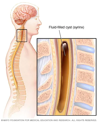
A syrinx is a fluid-filled neuroglial cavity within the spinal cord, in the brain stem, or in the nerves of the elbow
A syrinx is a rare, fluid-filled neuroglial cavity within the spinal cord (syringomyelia), in the brain stem (syringobulbia), or in the nerves of the elbow, usually in a young age.
Presentation
Symptoms usually begin insidiously between adolescence and age 45. Syringomyelia develops in the center of the spinal cord, causing a central cord syndrome. Pain and temperature sensory deficits occur early but may not be recognized for years. The first abnormality recognized may be a painless burn or cut. Syringomyelia typically causes weakness, atrophy, and often fasciculations and hyperreflexia of the hands and arms; a deficit in pain and temperature sensation in a capelike distribution over the shoulders, arms and back is characteristic. Light touch and position and vibration sensation are not affected. Later, spastic leg weakness develops. Deficits may be asymmetric.
Syringobulbia may cause vertigo, nystagmus, unilateral or bilateral loss of facial sensation, lingual atrophy and weakness, dysarthria, dysphagia, hoarseness, and sometimes peripheral sensory or motor deficits due to medullary compression.
Cause
A syrinx results when a watery, protective substance known as cerebrospinal fluid, that normally flows around the spinal cord and brain, transporting nutrients and waste products, collects in a small area of the spinal cord and forms a pseudocyst.
A number of medical conditions can cause an obstruction in the normal flow of cerebrospinal fluid, redirecting it into the spinal cord itself. For reasons that are only now becoming clear, this results in syrinx formation. Cerebrospinal fluid fills the syrinx. Pressure differences along the spine cause the fluid to move within the cyst. Physicians believe that it is this continual movement of fluid that results in cyst growth and further damage to the spinal cord.
In the case of syringomyelia, the syrinx can expand and elongate over time, destroying the spinal cord. Since the spinal cord connects the brain to nerves in the extremities, this damage may result in pain, weakness, and stiffness in the back, shoulders, arms, or legs. Other symptoms may include headaches and a loss of the ability to feel extremes of hot or cold, especially in the hands. Each patient experiences a different combination of symptoms. These symptoms typically vary depending on the extent and, often more critically, to the location of the syrinx within the spinal cord.
Syrinxes usually result from lesions that partially obstruct CSF flow. At least ½ of syrinxes occur in patients with congenital abnormalities of the craniocervical junction (e.g. herniation of cerebellar tissue into the spinal canal, called Chiari malformation), brain (e.g. encephalocele), or spinal cord (e.g. myelomeningocele—see Congenital Neurologic Anomalies: Brain Anomalies). For unknown reasons, these congenital abnormalities often expand during the teen or young adult years. A syrinx can also develop in patients who have a spinal cord tumor, scarring due to previous spinal trauma, or no known predisposing factors. About 30% of people with a spinal cord tumor eventually develop a syrinx.
- Tubbs RS, Elton S, Bartolucci AA, Grabb P, Oakes WJ (November 2000). “The position of the conus medullaris in children with a Chiari I malformation”. Pediatr Neurosurg. 33 (5): 249–251. doi:10.1159/000055963. PMID 11155061. S2CID
Chiari malformation is not always and perhaps not often “congenital.” Not only did nobody hear of Chiari until recently although they’ve been working on it for well over a hundred years (Chiari is named after Hans Chiari, a Viennese pathologist from the 19th century) and it has been featured on a plethora of medical television programing for the last 10-15 years, but they have been doing ultrasounds at all stages of pregnancy (for a very long time). The internet suggests ultrasound is sufficient for fetal diagnosis. So what’s up? When the condition is not present at birth, they call it acquired or secondary. The internet suggests Chiari malformation can be induced chemically and through trauma including birth injury and whiplash, among other things. Two types of Chiari malformation have been experimentally induced in pregnant hamsters (with a single dose of ‘vitamin A’ at a certain point of pregnancy) so I guess that would qualify as “congenital’ but it is still a poisoning. There may be some association with growth hormone (a couple of conflicting articles spurs that comment). It may be worth looking up in more detail.
- Marin-Padilla M, Marin-Padilla TM. Morphogenesis of experimentally induced Arnold–Chiari malformation. J Neurol Sci. 1981 Apr;50(1):29-55. doi: 10.1016/0022-510x(81)90040-x. PMID: 7229658. The administration of a single dose of vitamin A to pregnant hamsters, early during the morning of their 8th day of gestation, induces types I and II Arnold–Chiari malformation (ACM), as well as various types of axial skeletal-dysraphic disorders known to be associated with the human disease.
- Hida K, Iwasaki Y, Imamura H, Abe H. Birth injury as a causative factor of syringomyelia with Chiari type I deformity. J Neurol Neurosurg Psychiatry. 1994 Mar;57(3):373-4. doi: 10.1136/jnnp.57.3.373. PMID: 8158190; PMCID: PMC1072833.
- Massimi L, Della Pepa GM, Tamburrini G, Di Rocco C. Sudden onset of Chiari malformation Type I in previously asymptomatic patients. J Neurosurg Pediatr. 2011 Nov;8(5):438-42. doi: 10.3171/2011.8.PEDS11160. PMID: 22044365.
- Bunc G, Vorsic M. Presentation of a previously asymptomatic Chiari I malformation by a flexion injury to the neck. J Neurotrauma. 2001 Jun;18(6):645-8. doi: 10.1089/089771501750291882. PMID: 11437087.
- Uzoigwe CE, Shabani F, Chami G, El-Tayeb M. Simple whiplash? J Bone Joint Surg Br. 2009 Aug;91(8):1103-4. doi: 10.1302/0301-620X.91B8.22266. PMID: 19651845.
- O’Grady MJ, Cody D. Symptomatic Chiari 1 malformation after initiation of growth hormone therapy. J Pediatr. 2011 Apr;158(4):686. doi: 10.1016/j.jpeds.2010.09.079. Epub 2010 Nov 12. PMID: 21074170.
- William A Kerr & Jill E Hobbs (February 2002). “The North American-European Union Dispute Over Beef Produced Using Growth Hormones: A Major Test for the New International Trade Regime”. The World Economy. 25 (2): 283–296. doi:10.1111/1467-9701.00431. S2CID 154707486.
A wikidoc suggests Chiari affects more females than males (3:1) and it is no surprise that the same article says “The prevalence of Arnold Chiari malformation is unknown since most of the cases are accidentally found,” which, in case I didn’t say it clearly before, is completely unacceptable with ultrasounds up the kazoo and many of them all over social media. If that female/male ratio is correct, there could be estrogen factor or similar factors which we know are connected to growth hormone (although this is disputed?). A personal refresher because there are too many mentions of hormone and factor in addition to all manner of Chiari (why do they name these horrible problems after people like they just discovered gold? Hopefully not because that’s exactly what they did because something is not at all right with all this ‘healthcare’.) The things:
- Is growth factor the same as growth hormone?
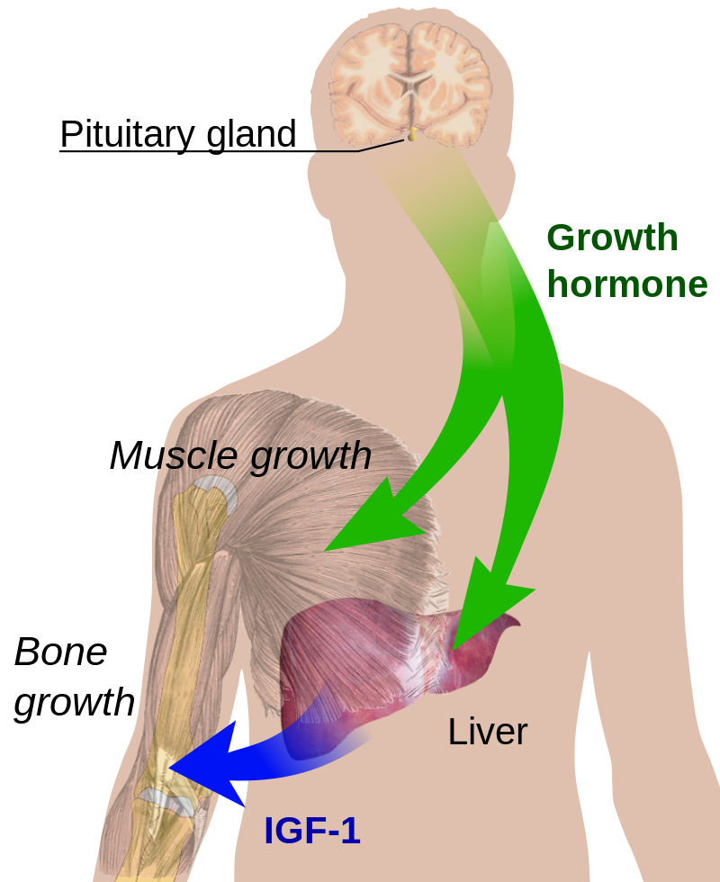
- Growth hormone (GH) or somatotropin, also known as human growth hormone (hGH or HGH) in its human form, is a peptide hormone that stimulates growth, cell reproduction, and cell regeneration in humans and other animals. It is thus important in human development. GH also stimulates production of IGF-1 and increases the concentration of glucose and free fatty acids. It is a type of mitogen which is specific only to the receptors on certain types of cells. GH is a 191-amino acid, single-chain polypeptide that is synthesized, stored and secreted by somatotropic cells within the lateral wings of the anterior pituitary gland. In its role as an anabolic agent, HGH has been used by competitors in sports since at least 1982, and has been banned by the IOC and NCAA. The names somatotropin (STH) or somatotropic hormone refer to the growth hormone produced naturally in animals and extracted from carcasses. Hormone extracted from human cadavers is abbreviated hGH. The main growth hormone produced by recombinant DNA technology has the approved generic name (INN) somatropin and the brand name Humatrope, and is properly abbreviated rhGH in the scientific literature. Since its introduction in 1992 Humatrope has been a banned sports doping agent, and in this context is referred to as HGH. The term growth hormone has been incorrectly applied to refer to anabolic sex hormones in the European beef hormone controversy, which initially restricts the use of estradiol, progesterone, testosterone, zeranol, melengestrol acetate and trenbolone acetate. In the United States, it is legal to give a bovine GH to dairy cows to increase milk production, and is legal to use GH in raising cows for beef; see article on Bovine somatotropin, cattle feeding, dairy farming and the beef hormone controversy. The use of GH in poultry farming is illegal in the United States. Similarly, no chicken meat for sale in Australia is administered hormones. Several companies have attempted to have a version of GH for use in pigs (porcine somatotropin) approved by the FDA but all applications have been withdrawn. In 1985, unusual cases of Creutzfeldt–Jakob disease were found in individuals that had received cadaver-derived HGH ten to fifteen years previously. Based on the assumption that infectious prions causing the disease were transferred along with the cadaver-derived HGH, cadaver-derived HGH was removed from the market. In 1985, biosynthetic human growth hormone replaced pituitary-derived human growth hormone for therapeutic use in the U.S. and elsewhere.
- Ranabir S, Reetu K (January 2011). “Stress and hormones”. Indian Journal of Endocrinology and Metabolism. 15 (1): 18–22. doi:10.4103/2230-8210.77573. PMC 3079864. PMID 21584161.
- Greenwood FC, Landon J (April 1966). “Growth hormone secretion in response to stress in man”. Nature. 210 (5035): 540–1. Bibcode:1966Natur.210..540G. doi:10.1038/210540a0. PMID 5960526. S2CID 1829264.
- Powers M (2005). “Performance-Enhancing Drugs”. In Leaver-Dunn D, Houglum J, Harrelson GL (eds.). Principles of Pharmacology for Athletic Trainers. Slack Incorporated. pp. 331–332. ISBN 978-1-55642-594-3.
- Daniels ME (1992). “Lilly’s Humatrope Experience”. Nature Biotechnology. 10 (7): 812. doi:10.1038/nbt0792-812a. S2CID 46453790.
- Saugy M, Robinson N, Saudan C, Baume N, Avois L, Mangin P (July 2006). “Human growth hormone doping in sport”. British Journal of Sports Medicine. 40 Suppl 1 (Suppl 1): i35–9. doi:10.1136/bjsm.2006.027573. PMC 2657499. PMID 16799101.
- William A Kerr & Jill E Hobbs (February 2002). “The North American-European Union Dispute Over Beef Produced Using Growth Hormones: A Major Test for the New International Trade Regime”. The World Economy. 25 (2): 283–296. doi:10.1111/1467-9701.00431. S2CID 154707486.
- “Chicken from Farm to Table | USDA Food Safety and Inspection Service”. Fsis.usda.gov. 2011-04-06. Archived from the original on 2011-09-03. Retrieved 2011-08-26.
- “Poultry Industry Frequently Asked Questions”. U.S. Poultry & Egg Association. Retrieved June 21, 2012.
- “Hormones”. Australian Chicken Meat Federation. Retrieved 20 June 2016.
- “Center for Veterinary Medicine Master” (PDF). www.fda.gov. 2011-04-06. Retrieved 2011-08-28.
- Maybe NG (1984). “Direct expression of human growth in Escherichia coli with the lipoprotein promoter”. In Bollon AP (ed.). Recombinant DNA products: insulin, interferon, and growth hormone. Boca Raton: CRC Press. ISBN 978-0-8493-5542-4.
- Beck JC, Mcgarry EE, Dyrenfurth I, Venning EH (May 1957). “Metabolic effects of human and monkey growth hormone in man”. Science. 125 (3253): 884–5. Bibcode:1957Sci…125..884B. doi:10.1126/science.125.3253.884. PMID 13421688.
- Beck JC, McGARRY EE, Dyrenfurth I, Venning EH (November 1958). “The metabolic effects of human and monkey growth hormone in man”. Annals of Internal Medicine. 49 (5): 1090–105. doi:10.
- Gardner DG, Shoback D (2007). Greenspan’s Basic and Clinical Endocrinology (8th ed.). New York: McGraw-Hill Medical. pp. 193–201. ISBN 978-0-07-144011-0.
- A growth factor is a naturally occurring substance capable of stimulating cell proliferation, wound healing, and occasionally cellular differentiation. Usually it is a secreted protein or a steroid hormone. Growth factors are important for regulating a variety of cellular processes. Growth factors typically act as signaling molecules between cells. Examples are cytokines and hormones that bind to specific receptors on the surface of their target cells. They often promote cell differentiation and maturation, which varies between growth factors. For example, epidermal growth factor (EGF) enhances osteogenic differentiation (osteogenesis or bone formation), while fibroblast growth factors and vascular endothelial growth factors stimulate blood vessel differentiation (angiogenesis).
- “growth factor” at Dorland’s Medical Dictionary
- Del Angel-Mosqueda C, Gutiérrez-Puente Y, López-Lozano AP, Romero-Zavaleta RE, Mendiola-Jiménez A, Medina-De la Garza CE, Márquez-M M, De la Garza-Ramos MA (September 2015). “Epidermal growth factor enhances osteogenic differentiation of dental pulp stem cells in vitro”. Head & Face Medicine. 11: 29. doi:10.1186/s13005-015-0086-5. PMC 4558932. PMID 26334535.

Estradiol (hormone or medication?) and Polyestradiol phosphate (which is not to be confused with Polyestriol phosphate or Estradiol phosphate) have been mentioned in the context of the disorder, as have others including prolactin.
- https://healthonecares.com/locations/colorado-chiari-institute/chiari-malformation-related-conditions.dot Increased secretion of prolactin (hyperprolactinemia) (11%) Empty Sella
I may be confusing/conflating Chiari disorders as:
Chiari syndrome or Chiari’s disease may refer to one of the following diseases named after the 19th century Austrian pathologist Hans Chiari:
- Arnold–Chiari malformation, or simply “Chiari malformation”, a malformation of the brain
- Budd–Chiari syndrome, a disease with typical symptoms of abdominal pain, ascites and hepatomegaly caused by occlusion of the hepatic veins (which may or may not be associated with pregnancy, postpartum period, oral contraceptives) and may progress to and/or involve encephalopathy.
- Chiari–Frommel syndrome, an older term for hyperprolactinaemia with extended postpartum galactorrhea and amenorrhea.
- “Chiari network” is ‘a remnant of the eustachian valve located in the right atrium. The attached articles says incomplete involution of the fetal sinus venosus valves results in “redundant” Chiari’s network, which may compromise cardiovascular function.
- Bendadi F, van Tijn DA, Pistorius L, Freund MW. Chiari’s network as a cause of fetal and neonatal pathology. Pediatr Cardiol. 2012 Jan;33(1):188-91. doi: 10.1007/s00246-011-0114-6. Epub 2011 Sep 10. PMID: 21909773; PMCID: PMC3248639. This report describes a case with the novel finding of prenatal compromise due to redundant Chiari’s network and an uncommon case with significant postnatal symptoms. In both cases, the symptoms (fetal hydrops and postnatal cyanosis) resolved spontaneously. The variety of cardiovascular pathologies described in the literature is believed to be associated with persistence of a Chiari network. That’s interesting because cyanosis is a symptom of other Chiari disorders and I’m going to look up that other word right now. Hydrops foetalis or hydrops fetalis is a condition in the fetus characterized by an accumulation of fluid, or edema, in at least two fetal compartments. That’s interesting because Rh disease is the only immune cause of hydrops fetalis. Hemolysis caused by the Rh incompatibility, causes extramedullary hematopoiesis in the fetal liver and bone marrow. The push to make more erythroblasts to help compensate with the hemolysis over works the liver causing hepatomegaly. The resulting liver dysfunction decreases albumin output which in turn decreases oncotic pressure. Consequentially, the decrease in pressure results in overall peripheral edema and ascites. They say that is currently not a main cause since preventative methods developed in the 1970s resulted in marked decline in Erythroblastosis fetalis, also known as Rh disease, There is a long list of nonimmune causes that may or may not be connected to fetal anemia.
- Jagannathan-Bogdan, Madhumita; Zon, Leonard I. (2013-06-15). “Hematopoiesis”. Development. 140 (12): 2463–2467. doi:10.1242/dev.083147. ISSN 0950-1991. PMC 3666375. PMID 23715539.

I have a minor interest in some of these things ever since I tried to amateurly (re)diagnose a long gone FDR who was disabled after he fell off a boat. (FDR was diagnosed with infantile paralysis, better known as polio, in 1921, at the age of 39.) It may have been the racoon eyes that set me off but that was years ago. I think I was looking at whiplash along with internal decapitation and several other things back then. If I saved the images, and I think I did, I may be able to sew that back together…as much as it ever was.
Back to Syringomyelia
Syringomyelia is a paramedian, usually irregular, longitudinal cavity.[citation needed] It most often affects the cervical and thoracic regions but may extend further down or up into the brainstem (syringobulbia). Syringobulbia, which is rare, usually occurs as a slitlike gap within the lower brain stem and may disrupt or compress the lower cranial nerves or ascending sensory or descending motor pathways.[citation needed]
- Eisen, Andrew. “Disorders affecting the spinal cord”. UpToDate. Retrieved 18 April 2017.
Diagnosis
A syrinx is suggested by an unexplained central cord syndrome or other characteristic neurologic deficits, particularly pain and temperature sensory deficits in a capelike distribution. MRI of the entire spinal cord and brain is done. Administration of intravenous Gadolinium during the MRI examination is useful for detecting any associated tumor.[citation needed]
Treatment
Underlying problems (e.g. craniocervical junction abnormalities, postoperative scarring, spinal tumors) are corrected when possible. Surgical decompression of the foramen magnum and upper cervical cord is the only useful treatment, but surgery usually cannot reverse severe neurologic deterioration.
Etymology
Syrinx is taken directly from the ancient Greek word for “tube.” It is the root of the word “syringe.”
- Le, Tao; Bhushan, Vikas; Vasan, Neil (2010). First Aid for the USMLE Step 1: 2010 20th Anniversary Edition. USA: The McGraw-Hill Companies, Inc. pp. 127. ISBN 978-0-07-163340-6.
- Harper, Douglas. “Etymology of syringe.” Online Etymology Dictionary, https://www.etymonline.com/word/syringe. Accessed 13 April, 2023.
References
- Tubbs RS, Elton S, Bartolucci AA, Grabb P, Oakes WJ (November 2000). “The position of the conus medullaris in children with a Chiari I malformation”. Pediatr Neurosurg. 33 (5): 249–251. doi:10.1159/000055963. PMID 11155061. S2CID 7270480.
- Eisen, Andrew. “Disorders affecting the spinal cord”. UpToDate. Retrieved 18 April 2017.
- Le, Tao; Bhushan, Vikas; Vasan, Neil (2010). First Aid for the USMLE Step 1: 2010 20th Anniversary Edition. USA: The McGraw-Hill Companies, Inc. pp. 127. ISBN 978-0-07-163340-6.
External links
ANEMIA, Body temperature, Brain Stem, Cerebrospinal Fluid, Chiari malformation, Cows, Craniocervical junction, Edema, Elbow, Estradiol, Estrogen, FDR, Foramen magnum, Growth factor, Growth hormone, Hormones, Monkey Business, Neuroglial, Pain, Periorbital ecchymosis, Poison, Polio, Prolactin, Pseudocyst, Racoon eyes, Rh, Spinal Cord, Syringe, Syrinx, Toxins


Leave a Reply