Cells in the APUD system may include Juxtaglomerular cells (JG cells), the renin producing cells in the kidney
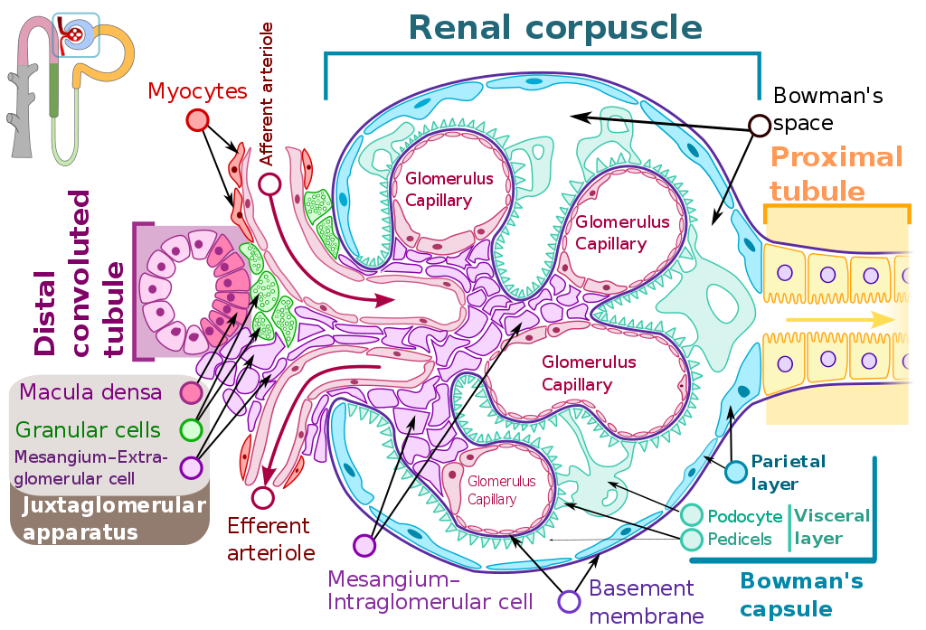
Drawing of renal corpuscle showing juxtaglomerular cells, macula densa cells and extraglomerular mesangium.
Juxtaglomerular cells (JG cells), also known as juxtaglomerular granular cells are cells in the kidney that synthesize, store, and secrete the enzyme renin.
Juxtaglomerular cells secrete renin in response to a drop in pressure detected by stretch receptors in the vascular walls, or when stimulated by macula densa cells. Macula densa cells are located in the distal convoluted tubule, and stimulate juxtaglomerular cells to release renin when they detect a drop in chloride concentration in tubular fluid. Together, juxtaglomerular cells, extraglomerular mesangial cells and macula densa cells comprise the juxtaglomerular apparatus.
In appropriately stained tissue sections, juxtaglomerular cells are distinguished by their granulated cytoplasm.
The juxtaglomerular cell is a cell that is located near the glomerulus, hence its name.
Similar to cardiac tissue, juxtaglomerular cells harbor β1 adrenergic receptors. When stimulated by epinephrine or norepinephrine, these receptors induce the secretion of renin. These cells also respond directly to a decrease in systemic blood pressure which is manifested as a lower renal perfusion pressure.
Morrison, Janna L.; Lumbers, Eugenie; Bennet, Laura; Black, Jane (November 2013). “Introduction: Celebrating Emeritus Scientia Professor Eugenie R Lumbers AM and Professor Caroline McMillen”. Clinical and Experimental Pharmacology and Physiology. 40 (11): 740–742. doi:10.1111/1440-1681.12180. PMID24117727. S2CID44555887.
Lumbers, ER (30 June 1971). “Activation of renin in human amniotic fluid by low pH”. Enzymologia. 40 (6): 329–336. PMID4105386.
Synthesis
In addition to juxtaglomerular cells, prorenin is also synthesised by other organs, such as the adrenal glands, the ovaries, the testis and the pituitary gland, which is why it is found in the plasma of anephric individuals.
Blood concentration levels of prorenin are between 5 and 10 times higher than those of renin. There is evidence to suggest that, in diabetes mellitus, prorenin levels are even higher. One study using relatively newer technology found that blood concentrations levels may be several order of magnitude higher than previously believed, and placing it at micrograms rather than nanograms per millilitre.
Fujimoto, Kazumi; Kawamura, Sayuki; Bando, Satoru; Kamata, Yuji; Kodera, Yoshio; Shichiri, Masayoshi (June 2021). “Circulating prorenin: its molecular forms and plasma concentrations”. Hypertension Research. 44 (6): 674–684. doi:10.1038/s41440-020-00610-0. PMID33564180. S2CID231859379.
Pregnancy
Prorenin occurs in very high concentrations in amniotic fluid and amnion. It is secreted in large amounts from the placenta and womb, and from the ovaries.
There is no evidence that prorenin can be converted into renin in the circulation. Therefore, the granular (JG) cells seem to be the only source of active renin.
Morrison, Janna L.; Lumbers, Eugenie; Bennet, Laura; Black, Jane (November 2013). “Introduction: Celebrating Emeritus Scientia Professor Eugenie R Lumbers AM and Professor Caroline McMillen”. Clinical and Experimental Pharmacology and Physiology. 40 (11): 740–742. doi:10.1111/1440-1681.12180. PMID24117727. S2CID44555887.
Lumbers, ER (30 June 1971). “Activation of renin in human amniotic fluid by low pH”. Enzymologia. 40 (6): 329–336. PMID4105386.
Leave a Reply