Parafollicular cells aka C cells secrete calcitonin and several neuroendocrine peptides
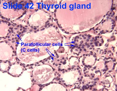
Parafollicular cells, also called C cells, are neuroendocrine cells in the thyroid. The primary function of these cells is to secrete calcitonin. They are located adjacent to the thyroid follicles and reside in the connective tissue. These cells are large and have a pale stain compared with the follicular cells. In teleost and avian species these cells occupy a structure outside the thyroid gland named the ultimobranchial body.
Structure
Parafollicular cells are pale-staining cells found in small number in the thyroid and are typically situated basally in the epithelium, without direct contact with the follicular lumen. They are always situated within the basement membrane, which surrounds the entire follicle.
Development
Parafollicular cells are derived from pharyngeal endoderm.
Johansson, E., Andersson, L., Örnros, J., Carlsson, T., Ingeson-Carlsson, C., Liang, S., … Nilsson, M. (2015). Revising the embryonic origin of thyroid C cells in mice and humans. Development, 142(20), 3519–3528. http://doi.org/10.1242/dev.126581
Embryologically, they associate with the ultimobranchial body, which is a ventral derivative of the fourth (or fifth) pharyngeal pouch. Parafollicular cells were previously believed to be derived from the neural crest based on a series of experiments in quail-chick chimeras.
Le Douarin N, Fontaine J, Le Lièvre C (March 1974). “New studies on the neural crest origin of the avian ultimobranchial glandular cells–interspecific combinations and cytochemical characterization of C cells based on the uptake of biogenic amine precursors”. Histochemistry. 38 (4): 297–305. doi:10.1007/bf00496718. PMID4135055. S2CID7551942.
Parafollicular cells secrete calcitonin, a hormone that participates in the regulation of calcium metabolism. Calcitonin lowers blood levels of calcium by inhibiting the resorption of bone by osteoclasts, and its secretion is increased proportionally with the concentration of calcium.
Melmed S, Polonsky KS, Larsen PR, Kronenberg HM (2011). Williams Textbook of Endocrinology (12th ed.). Saunders. pp. 1250–1252. ISBN978-1437703245.
Parafollicular cells are also known to secrete in smaller quantities several neuroendocrine peptides such as serotonin, somatostatin or CGRP.
Zabel M (December 1984). “Ultrastructural localization of calcitonin, somatostatin and serotonin in parafollicular cells of rat thyroid”. The Histochemical Journal. 16 (12): 1265–72. doi:10.1007/bf01003725. PMID6152264. S2CID7889687.
Johansson, E., Andersson, L., Örnros, J., Carlsson, T., Ingeson-Carlsson, C., Liang, S., … Nilsson, M. (2015). Revising the embryonic origin of thyroid C cells in mice and humans. Development, 142(20), 3519–3528. http://doi.org/10.1242/dev.126581
Le Douarin N, Fontaine J, Le Lièvre C (March 1974). “New studies on the neural crest origin of the avian ultimobranchial glandular cells–interspecific combinations and cytochemical characterization of C cells based on the uptake of biogenic amine precursors”. Histochemistry. 38 (4): 297–305. doi:10.1007/bf00496718. PMID4135055. S2CID7551942.
Melmed S, Polonsky KS, Larsen PR, Kronenberg HM (2011). Williams Textbook of Endocrinology (12th ed.). Saunders. pp. 1250–1252. ISBN978-1437703245.
Zabel M (December 1984). “Ultrastructural localization of calcitonin, somatostatin and serotonin in parafollicular cells of rat thyroid”. The Histochemical Journal. 16 (12): 1265–72. doi:10.1007/bf01003725. PMID6152264. S2CID7889687.
Kameda Y (October 1987). “Localization of immunoreactive calcitonin gene-related peptide in thyroid C cells from various mammalian species”. The Anatomical Record. 219 (2): 204–12. doi:10.1002/ar.1092190214. PMID3120623. S2CID12517073.
Leave a Reply