Hitt, J. C.; Bryon, D. M.; Modianos, D. T. (1973). “Effects of rostral medial forebrain bundle and olfactory tubercle lesions upon sexual behavior of male rats”. Journal of Comparative and Physiological Psychology. 82 (1): 30–36. doi:10.1037/h0033797. PMID4567890.
Koob, G. F.; Riley, S. J.; Smith, S. C.; Robbins, T. W. (1978). “Effects of 6-hydroxydopamine lesions of the nucleus accumbens septi and olfactory tubercle on feeding, locomotor activity, and amphetamine anorexia in the rat”. Journal of Comparative and Physiological Psychology. 92 (5): 917–927. doi:10.1037/h0077542. PMID282297.
The OT is interconnected with numerous brain regions, especially the sensory, arousal, and reward centers, thus making it a potentially critical interface between processing of sensory information and the subsequent behavioral responses.
The OT is a composite structure that receives direct input from the olfactory bulb and contains the morphological and histochemical characteristics of the ventral pallidum and the striatum of the forebrain.
Heimer, L.; Wilson, R. D. (1975). “The subcortical projections of the allocortex: Similarities in the neural connections of the hippocampus, the piriform cortex and the neocortex”. In Santini, Maurizio (ed.). Golgi Centennial Symposium Proceedings. New York: Raven. pp. 177–193. ISBN978-0911216806.
Ikemoto S (2010). “Brain reward circuitry beyond the mesolimbic dopamine system: a neurobiological theory”. Neuroscience & Biobehavioral Reviews. 35 (2): 129–50. doi:10.1016/j.neubiorev.2010.02.001. PMC2894302. PMID20149820. Recent studies on intracranial self-administration of neurochemicals (drugs) found that rats learn to self-administer various drugs into the mesolimbic dopamine structures–the posterior ventral tegmental area, medial shell nucleus accumbens and medial olfactory tubercle. … In the 1970s it was recognized that the olfactory tubercle contains a striatal component, which is filled with GABAergic medium spiny neurons receiving glutamatergic inputs form cortical regions and dopaminergic inputs from the VTA and projecting to the ventral pallidum just like the nucleus accumbens Figure 3: The ventral striatum and self-administration of amphetamine
Brunton, Laurence L.; Hilal-Dandan, Randa; Knollmann, Bjorn C. (2018). Goodman & Gilman’s – The pharmacological basis of therapeutics. Mc Graw Hill Education. p. 329. ISBN978-1-25-958473-2.
Structure
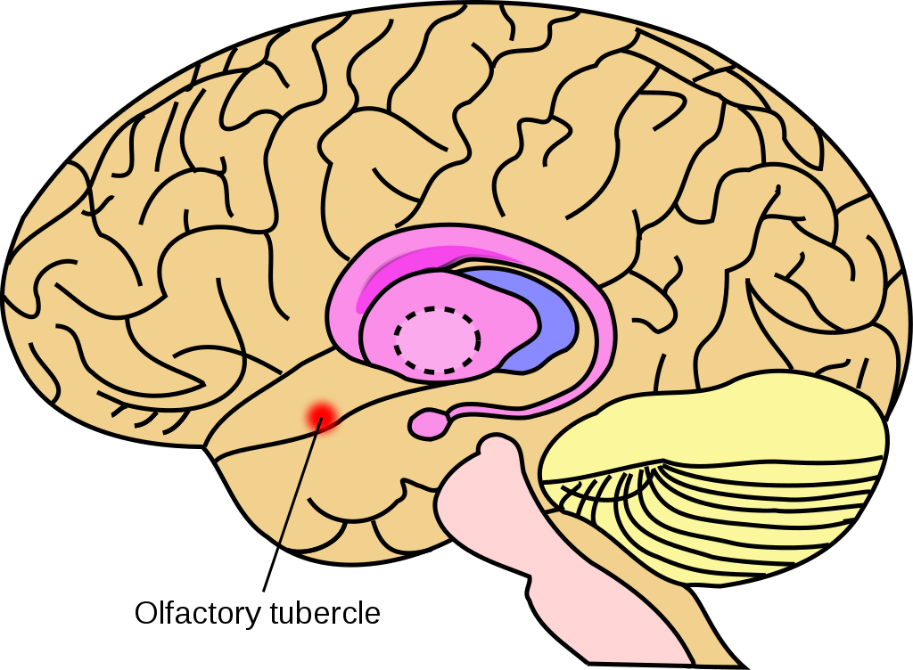
The olfactory tubercle differs in location and relative size between humans, other primates, rodents, birds, and other animals. In most cases, the olfactory tubercle is identified as a round bulge along the basal forebrain anterior to the optic chiasm and posterior to the olfactory peduncle.
In humans and other primates, visual identification of the olfactory tubercle is not easy because the basal forebrain bulge is small in these animals.
Millhouse, O. E.; Heimer, L. (1984). “Cell configurations in the olfactory tubercle of the rat”. The Journal of Comparative Neurology. 228 (4): 571–597. doi:10.1002/cne.902280409. PMID6490970. S2CID9261393.
With regard to functional anatomy, the olfactory tubercle can be considered to be a part of three larger networks. First, it is considered to be part of the basal forebrain, the nucleus accumbens, and the amygdaloid nuclei because of its location along the rostral ventral region of the brain, that is, the front-bottom part. Second, it is considered to be part of the olfactory cortex because it receives direct input from the olfactory bulb. Third, it is also considered to be part of the ventral striatum based on anatomy, neurochemical, and embryology data.
One of the most striking features of the olfactory tubercle is the closely packed crescent-shape cell clusters, which are located mostly in layer III and sometimes in layer II. These cell clusters, called the islands of Calleja, are innervated by dopaminergic projections from the nucleus accumbens and the substantia nigra, suggesting the role that the olfactory tubercle plays in the reward system.
The olfactory tubercle is a multi-sensory processing center due to the number of innervations going to and from other brain regions such as the amygdala, thalamus, hypothalamus, hippocampus, brain stem, auditory and visual sensory fibers, and a number of structures in the reward–arousal system, as well as the olfactory cortex. Due to its many innervations from other brain regions, the olfactory tubercle is involved in merging information across the senses, such as olfactory/audition and olfactory/visual integrations, possibly in a behaviorally relevant manner. Thus, damage to the olfactory tubercle is likely to affect the functionality of all these areas of the brain. Examples of such disruption include changes in normal odor-guided behavior, and impairments in modulating state and motivational behavior, which are common in psychiatric disorders such as schizophrenia, dementia and depression.
Rupp, C. I.; Fleischhacker, W. W.; Kemmler, G.; Kremser, C.; Bilder, R. M.; Mechtcheriakov, S.; Szeszko, P. R. (2005). “Olfactory functions and volumetric measures of orbitofrontal and limbic regions in schizophrenia”. Schizophrenia Research. 74 (2–3): 149–161. doi:10.1016/j.schres.2004.07.010. PMID15721995. S2CID11026266.
Murphy, C.; Nordin, S.; Jinich, S. (1999). “Very early decline in recognition memory for odors in Alzheimer’s disease”. Aging, Neuropsychology, and Cognition. 6 (3): 229–240. doi:10.1076/anec.6.3.229.777.
Negoias, S.; Croy, I.; Gerber, J.; Puschmann, S.; Petrowski, K.; Joraschky, P.; Hummel, T. (2010). “Reduced olfactory bulb volume and olfactory sensitivity in patients with acute major depression”. Neuroscience. 169 (1): 415–421. doi:10.1016/j.neuroscience.2010.05.012. PMID20472036. S2CID207248484.
The olfactory tubercle has been shown to play a large role in behavior. Unilateral lesions in the olfactory tubercle have been shown to alter attention, social and sensory responsiveness, and even locomotor behavior.
Koob, G. F.; Riley, S. J.; Smith, S. C.; Robbins, T. W. (1978). “Effects of 6-hydroxydopamine lesions of the nucleus accumbens septi and olfactory tubercle on feeding, locomotor activity, and amphetamine anorexia in the rat”. Journal of Comparative and Physiological Psychology. 92 (5): 917–927. doi:10.1037/h0077542. PMID282297.
Bilateral lesions have been shown to reduce copulatory behavior in male rats. The olfactory tubercle has also been shown to be especially involved in reward and addictive behaviors. Rats have been shown to administer cocaine into the olfactory tubercle more than the nucleus accumbens and ventral pallidum, other reward centers in the brain. In fact, they will administer cocaine into the olfactory tubercle at about 200 times per hour and even till death.
Functional contributions of the olfactory tubercle to olfaction are currently unclear; however, there is evidence of a perceptual role that it may play. Work from Zelano, et al. suggest that the olfactory tubercle may be crucial in sorting out the sources of olfactory information.
Zelano, C.; Montag, J.; Johnson, B.; Khan, R.; Sobel, N. (2007). “Dissociated representations of irritation and valence in human primary olfactory cortex”. Journal of Neurophysiology. 97 (3): 1969–1976. doi:10.1152/jn.01122.2006. PMID17215504.
This suggests that it may also play a role in odor guided behavior. Thus, it may link perception of odor with action through its connections with attention, reward, and motivation systems of the basal forebrain.
Functional imaging data from this same group also shows that the olfactory tubercle is highly activated during tasks that engage attention, thus playing a large role in arousal-related systems.
Because the olfactory tubercle is a component of the ventral striatum, it is heavily interconnected with several affective-, reward-, and motivation-related centers of the brain. It also sits at the interface between the olfactory sensory input and state-dependent behavioral modulatory circuits, that is the area that modulates behavior during certain physiological and mental states. Thus, the olfactory tubercle may also play an important role in the mediation of odor approach and odor avoidance behavior, probably in a state-dependent manner.
Gervais, G. (1979). “Unilateral lesions of the olfactory tubercle modifying general arousal effects in the rat olfactory bulb”. Electroencephalography and Clinical Neurophysiology. 46 (6): 665–674. doi:10.1016/0013-4694(79)90104-4. PMID87311.
Anatomy
In general, the olfactory tubercle is located at the basal forebrain of the animal within the medial temporal lobe. Specifically, parts of the tubercle are included in the olfactory cortex and nested between the optic chiasm and olfactory tract and ventral to the nucleus accumbens. The olfactory tubercle consists of three layers, a molecular layer (layer I), the dense cell layer (layer II), and the multiform layer (layer III).
Millhouse, O. E.; Heimer, L. (1984). “Cell configurations in the olfactory tubercle of the rat”. The Journal of Comparative Neurology. 228 (4): 571–597. doi:10.1002/cne.902280409. PMID6490970. S2CID9261393.
Other than the islands of Calleja, which are characteristic of the tubercle, it is also noted for the being innervated by dopaminergic neurons from the ventral tegmental area. The olfactory tubercle also consists of heterogeneous elements, such as medial forebrain bundle, and has a ventral extension of the striatal complex. During the 1970s, the tubercle was found to contain a striatal component which is composed of GABAergicmedium spiny neurons.
Ikemoto S (2010). “Brain reward circuitry beyond the mesolimbic dopamine system: a neurobiological theory”. Neuroscience & Biobehavioral Reviews. 35 (2): 129–50. doi:10.1016/j.neubiorev.2010.02.001. PMC2894302. PMID20149820. Recent studies on intracranial self-administration of neurochemicals (drugs) found that rats learn to self-administer various drugs into the mesolimbic dopamine structures–the posterior ventral tegmental area, medial shell nucleus accumbens and medial olfactory tubercle. … In the 1970s it was recognized that the olfactory tubercle contains a striatal component, which is filled with GABAergic medium spiny neurons receiving glutamatergic inputs form cortical regions and dopaminergic inputs from the VTA and projecting to the ventral pallidum just like the nucleus accumbensFigure 3: The ventral striatum and self-administration of amphetamine
Calleja, C. (1893). La region olfactoria del cerebro. Madrid: Nicolas Moya.
The GABAergic neurons project to the ventral pallidum and receive glutamatergic inputs from cortical regions and dopaminergic inputs from the ventral tegmental area.
Meyer, G.; Gonzalez-Hernandez, T.; Carrillo-Padilla, F.; Ferres-Torres, R. (1989). “Aggregations of granule cells in the basal forebrain (islands of Calleja): Golgi and cytoarchitectonic study in different mammals, including man”. The Journal of Comparative Neurology. 284 (3): 405–428. doi:10.1002/cne.902840308. PMID2474005. S2CID29824764.
Millhouse, O. E. (1987). “Granule cells of the olfactory tubercle and the question of the islands of calleja”. The Journal of Comparative Neurology. 265 (1): 1–24. doi:10.1002/cne.902650102. PMID3693600. S2CID21826194
Morphological and neurochemical features
The ventral portion of the olfactory tubercle consists of three layers, whereas the dorsal portion contains dense cell clusters and adjoins the ventral pallidum (within the basal ganglia). The structure of the most ventral and anterior parts of the tubercle can be defined as anatomically defined hills (consisting of gyri and sulci) and clusters of cells.
The most common cell types in the olfactory tubercle are medium-size dense spine cells found predominantly in layer II (dense cell layer). The dendrites of these cells are covered by substance p immunoreactive (S.P.I) axons up into layer III (multiform layer).
Millhouse, O. E.; Heimer, L. (1984). “Cell configurations in the olfactory tubercle of the rat”. The Journal of Comparative Neurology. 228 (4): 571–597. doi:10.1002/cne.902280409. PMID6490970. S2CID9261393.
These cells also project into the nucleus accumbens and caudate putamen, thus linking the olfactory tubercle with the pallidum.
Fallon, JH. (Jun 1983). “The islands of Calleja complex of rat basal forebrain II: connections of medium and large sized cells”.Brain Research Bulletin. 10 (6): 775–93. doi:10.1016/0361-9230(83)90210-1. PMID6616269. S2CID4723010.
Other medium-size cells reside in layers II and III of the olfactory tubercle as well. These include the spine-poor neurons and spindle cells and they differ from the medium-size dense spine cells because they have sparse dendritic trees. The largest cells, and most striking feature of the olfactory tubercle, are densely packed crescent-shape cell clusters, Islands of Calleja that reside mostly in the dorsal portion of the olfactory tubercle, layer III, and can also be found in layer II. The olfactory tubercle also contains three classes of small cells found mostly in layers I and II. The first are pial cells (named as such because of location near pial surface), which look like miniature medium-size dense spine cells. The second are radiate cells and are easily identified by numerous multi-directional spineless dendrites. The third, small spine cells, are similar to the pial cells in that they also look like medium-size spine cells except they are not located near the pial surface.
Ribak, CE.; Fallon, JH. (Mar 1982). “The island of Calleja complex of rat basal forebrain. I. Light and electron microscopic observations”. The Journal of Comparative Neurology. 205 (3): 207–18. doi:10.1002/cne.902050302. PMID7076893. S2CID44954144
Development
Migrating cells from several developmental sites come together to form the olfactory tubercle. This includes the ventral ganglionic eminence (found in ventral part of telencephalon, where they form bulges in the ventricles that later become the basal ganglia, present only in embryonic stages) and the rostromedial telencephalic wall (of the forebrain).
Olfactory tubercle neurons originate as early as embryonic day 13 (E13), and the cell development occurs in a layer specific manner. The emergence of the three main layers of the olfactory tubercle begins almost simultaneously. The large neurons in layer III originate from E13 to E16, while the small and medium originate between E15 and E20. Like the small and medium cells in layer III, the cells of layer II and the striatal bridges also originate between E15 and E20 and develop in a lateral to medial gradient.
The granule cells of the islands of calleja originate between E19 and E22 and continue to migrate into the islands until long after birth.
Bedard, A.; Levesque, M.; Bernier, P. J.; Parent, A. (2002). “The rostral migratory stream in adult squirrel monkeys: contribution of new neurons to the olfactory tubercle and involvement of the antiapoptotic protein bcl-2”. European Journal of Neuroscience. 16 (10): 1917–1924. doi:10.1046/j.1460-9568.2002.02263.x. PMID12453055. S2CID31096044.
^ De Marchis, S.; Fasolo, A.; Puche, AC. (Aug 2004). “Subventricular zone-derived neuronal progenitors migrate into the subcortical forebrain of postnatal mice”. The Journal of Comparative Neurology. 476 (3): 290–300. doi:10.1002/cne.20217. PMID15269971. S2CID25911933.
Fibers from the lateral olfactory tract begin branching into the olfactory tubercle around E17. The lateral portion of the olfactory tubercle (which adjoins the olfactory tract) receives the densest fiber input and the medial portion receives light fiber projections. This branching continues until completion about the end of the first week after birth.
Schwob, JE.; Price, JL. (Feb 1984). “The development of axonal connections in the central olfactory system of rats”. The Journal of Comparative Neurology. 223 (2): 177–202. doi:10.1002/cne.902230204. PMID6200518. S2CID25870173
Deadwyler, S. A.; Foster, T. C.; Hampson, R. E. (1987). “Processing of sensory information in the hippocampus”. CRC Critical Reviews in Clinical Neurobiology. 2 (4): 335–355. PMID3297494.
This convergence has been shown to cause the perception of sound, caused by the interaction between smell and sound. This possibility has been supported by work from where olfactory tubercle displayed olfactory–auditory convergence.
Retinal projections have also been found in layer II of the olfactory tubercle, suggesting that it constitutes a region of olfactory and visual convergence.
Mick, G.; Cooper, H.; Magnin, M. (1993). “Retinal projection to the olfactory tubercle and basal telencephalon in primates”. The Journal of Comparative Neurology. 327 (2): 205–219. doi:10.1002/cne.903270204. PMID8425942. S2CID21784363
These visual sensory fibers arrive from the retinal ganglion cells. Thus, the olfactory tubercle may play a role in the perception of odors when a visual source is identified.
As far as olfaction is concerned, in vitro data from some studies suggest that the olfactory tubercle units have the functional capability of other olfactory center neurons in processing odor. It has been suggested that the olfactory tubercle may be crucial in determining the source of olfactory information and responds to odor inhalations that are attended to.
Zelano, C.; Montag, J.; Johnson, B.; Khan, R.; Sobel, N. (2007). “Dissociated representations of irritation and valence in human primary olfactory cortex”. Journal of Neurophysiology. 97 (3): 1969–1976. doi:10.1152/jn.01122.2006. PMID17215504
Role in behavior
The olfactory tubercle has been shown to be concerned primarily with the reception of sensory impulses from olfactory receptors.
Adey, W. R. (1959). “CHAPTER XXI”. In J. Field (ed.). The sense of smell. Handbook of physiology. Vol. I. Washington, D. C.: American Physiological Assn. pp. 535–548. Retrieved 2013-11-06.
Because of its connections to regions like the amygdala and hippocampus, the olfactory tubercle may play a role in behavior. Rats rely heavily on olfactory sensory input from olfactory receptors for behavioral attitudes.
Barnett, S A (1963). The rat; a study in behaviour. Chicago: Aldine Pub. Co. OCLC558946.
Studies show that bilateral lesions in the olfactory tubercle significantly reduce stereotyped behavior such as copulatory behavior in male rats and a reduction in sniffing and chewing behaviors.
Koob, G. F.; Riley, S. J.; Smith, S. C.; Robbins, T. W. (1978). “Effects of 6-hydroxydopamine lesions of the nucleus accumbens septi and olfactory tubercle on feeding, locomotor activity, and amphetamine anorexia in the rat”. Journal of Comparative and Physiological Psychology. 92 (5): 917–927. doi:10.1037/h0077542. PMID282297.
Asher, I. M.; Aghajanian, G. K. (1974). “6-hydroxydopamine lesions of olfactory tubercles and caudate nuclei: Effect on amphetamine-induced stereo-typed behavior in rats”. Brain Research. 82 (1): 1–12. doi:10.1016/0006-8993(74)90888-9. PMID4373138.
McKenzie, GM. (1972). “Role of the tuberculum olfactorium in streotyped behaviour induced by apomorphine in the rat”. Psychopharmacologia. 23 (3): 212–9. doi:10.1007/bf00404127. PMID5026945. S2CID6928275.
These stereotyped inhibitions may have been caused by the removal of central neuronal processes other than the dopaminergic cells in the olfactory tubercle. Unilateral lesions have been shown to alter attention, social and sensory responsiveness, and even locomotor behavior in rats.
Koob, G. F.; Riley, S. J.; Smith, S. C.; Robbins, T. W. (1978). “Effects of 6-hydroxydopamine lesions of the nucleus accumbens septi and olfactory tubercle on feeding, locomotor activity, and amphetamine anorexia in the rat”. Journal of Comparative and Physiological Psychology. 92 (5): 917–927. doi:10.1037/h0077542. PMID282297.
Arousal and reward
The dopaminergic neurons from the ventral tegmental area that innervate the olfactory tubercle enable the tubercle to play roles in reward and arousal and appears to partially mediate cocaine reinforcement.
The anteromedial portions of the tubercle have been shown to mediate some of the rewarding effects of drugs like cocaine and amphetamine. This has been shown in studies where rats learn to self-administer cocaine at significantly high rates into the tubercle. Injections of cocaine into the tubercle induce robust locomotion and rearing behavior in rats.
The multi-sensory nature of the olfactory tubercle and the many innervations it receives from other brain regions, especially the direct input from the olfactory bulb and innervations from the ventral tegmental area, makes it likely to be involved in several psychiatric disorders in which olfaction and dopamine receptors are affected. Many studies have found reduced olfactory sensitivity in patients with major depressive disorders (MDD) and dementia and schizophrenia. Patients with MDD have been shown to have reduced olfactory bulb and olfactory cortex as compared to normal people.
Negoias, S.; Croy, I.; Gerber, J.; Puschmann, S.; Petrowski, K.; Joraschky, P.; Hummel, T. (2010). “Reduced olfactory bulb volume and olfactory sensitivity in patients with acute major depression”. Neuroscience. 169 (1): 415–421. doi:10.1016/j.neuroscience.2010.05.012. PMID20472036. S2CID207248484.
In dementias, especially of the Alzheimer’s disease type, the olfactory bulb, anterior olfactory nucleus, and orbitofrontal cortex, all areas of the brain that process olfaction are affected. The deficits observed in dementia include decrease in odor threshold sensitivity, odor identification and odor memory.
Nordin, S.; Murphy, C. (1996). “Impaired sensory and cognitive olfactory function in questionable Alzheimer’s disease”. Neuropsychology. 10 (1): 112–119. doi:10.1037/0894-4105.10.1.113.
Doty, R. L.; Perl, D. P.; Steele, J. C.; Chen, K. M.; Pierce, J. D. Jr.; Reyes, P.; Kurland, L. T. (1991). “Odor identification deficit of the parkinsonism-dementia complex of Guam: equivalence to that of Alzheimer’s and idiopathic Parkinson’s disease”. Neurology. 41 (5 Suppl 2): 77–80, discussion 80–81. doi:10.1212/WNL.41.5_Suppl_2.77. PMID2041598. S2CID36051446.
Murphy, C.; Nordin, S.; Jinich, S. (1999). “Very early decline in recognition memory for odors in Alzheimer’s disease”. Aging, Neuropsychology, and Cognition. 6 (3): 229–240. doi:10.1076/anec.6.3.229.777.
Patients with schizophrenia exhibit deficits in olfactory discrimination that are not seen in patients with other psychiatric disorders not mentioned here. Rupp, et al. found that in patients with schizophrenia olfactory sensitivity and discrimination as well as higher order identification abilities are reduced.
Rupp, C. I.; Fleischhacker, W. W.; Kemmler, G.; Kremser, C.; Bilder, R. M.; Mechtcheriakov, S.; Szeszko, P. R. (2005). “Olfactory functions and volumetric measures of orbitofrontal and limbic regions in schizophrenia”. Schizophrenia Research. 74 (2–3): 149–161. doi:10.1016/j.schres.2004.07.010. PMID15721995. S2CID11026266
As mentioned earlier, the olfactory tubercle may be involved in the perception of odors due to the inputs received from the bulb and thus, by extension, may play a role in these psychiatric disorders.
History
The olfactory tubercle was first described by Albert von Kölliker in 1896, who studied them in rats. Since then, there have been several histological and histochemical studies; done in this area to identify it in other rodents, cats, humans, non-human primates, and other species.
Koob, G. F.; Riley, S. J.; Smith, S. C.; Robbins, T. W. (1978). “Effects of 6-hydroxydopamine lesions of the nucleus accumbens septi and olfactory tubercle on feeding, locomotor activity, and amphetamine anorexia in the rat”. Journal of Comparative and Physiological Psychology. 92 (5): 917–927. doi:10.1037/h0077542. PMID282297.
Millhouse, O. E.; Heimer, L. (1984). “Cell configurations in the olfactory tubercle of the rat”. The Journal of Comparative Neurology. 228 (4): 571–597. doi:10.1002/cne.902280409. PMID6490970. S2CID9261393
Similar studies were done by several authors to find the cell composition and innervations to and from other regions in the OT. Over the years, several other methods have been employed to find the possible functions and role of the OT in the brain. These began with lesion studies and early electrophysiological recordings.
Koob, G. F.; Riley, S. J.; Smith, S. C.; Robbins, T. W. (1978). “Effects of 6-hydroxydopamine lesions of the nucleus accumbens septi and olfactory tubercle on feeding, locomotor activity, and amphetamine anorexia in the rat”. Journal of Comparative and Physiological Psychology. 92 (5): 917–927. doi:10.1037/h0077542. PMID282297.
Détári, L.; Juhász, G.; Kukorelli, T. (1984). “Firing properties of cat basal forebrain neurones during sleep-wakefulness cycle”. Electroencephalography and Clinical Neurophysiology. 58 (4): 362–368. doi:10.1016/0013-4694(84)90062-2. ISSN0013-4694. PMID6207005.
Suaud-Chagny, M.F.; Ponec, J.; Gonon, F. (1991). “Presynaptic autoinhibition of the electrically evoked dopamine release studied in the rat olfactory tubercle byin vivo electrochemistry”. Neuroscience. 45 (3): 641–652. doi:10.1016/0306-4522(91)90277-U. ISSN0306-4522. PMID1775239. S2CID46471029.
Gervais, G. (1979). “Unilateral lesions of the olfactory tubercle modifying general arousal effects in the rat olfactory bulb”. Electroencephalography and Clinical Neurophysiology. 46 (6): 665–674. doi:10.1016/0013-4694(79)90104-4. PMID87311.
Asher, I. M.; Aghajanian, G. K. (1974). “6-hydroxydopamine lesions of olfactory tubercles and caudate nuclei: Effect on amphetamine-induced stereo-typed behavior in rats”. Brain Research. 82 (1): 1–12. doi:10.1016/0006-8993(74)90888-9. PMID4373138.
Improvements in technology have made it possible to now place multiple electrodes in the olfactory tubercle and record from anesthetized and even awake animals participating in behavioral tasks.
Doty, R. L.; Perl, D. P.; Steele, J. C.; Chen, K. M.; Pierce, J. D. Jr.; Reyes, P.; Kurland, L. T. (1991). “Odor identification deficit of the parkinsonism-dementia complex of Guam: equivalence to that of Alzheimer’s and idiopathic Parkinson’s disease”. Neurology. 41 (5 Suppl 2): 77–80, discussion 80–81. doi:10.1212/WNL.41.5_Suppl_2.77. PMID2041598. S2CID36051446.
Hitt, J. C.; Bryon, D. M.; Modianos, D. T. (1973). “Effects of rostral medial forebrain bundle and olfactory tubercle lesions upon sexual behavior of male rats”. Journal of Comparative and Physiological Psychology. 82 (1): 30–36. doi:10.1037/h0033797. PMID4567890.
Koob, G. F.; Riley, S. J.; Smith, S. C.; Robbins, T. W. (1978). “Effects of 6-hydroxydopamine lesions of the nucleus accumbens septi and olfactory tubercle on feeding, locomotor activity, and amphetamine anorexia in the rat”. Journal of Comparative and Physiological Psychology. 92 (5): 917–927. doi:10.1037/h0077542. PMID282297.
Heimer, L.; Wilson, R. D. (1975). “The subcortical projections of the allocortex: Similarities in the neural connections of the hippocampus, the piriform cortex and the neocortex”. In Santini, Maurizio (ed.). Golgi Centennial Symposium Proceedings. New York: Raven. pp. 177–193. ISBN978-0911216806.
Ikemoto S (2010). “Brain reward circuitry beyond the mesolimbic dopamine system: a neurobiological theory”. Neuroscience & Biobehavioral Reviews. 35 (2): 129–50. doi:10.1016/j.neubiorev.2010.02.001. PMC2894302. PMID20149820. Recent studies on intracranial self-administration of neurochemicals (drugs) found that rats learn to self-administer various drugs into the mesolimbic dopamine structures–the posterior ventral tegmental area, medial shell nucleus accumbens and medial olfactory tubercle. … In the 1970s it was recognized that the olfactory tubercle contains a striatal component, which is filled with GABAergic medium spiny neurons receiving glutamatergic inputs form cortical regions and dopaminergic inputs from the VTA and projecting to the ventral pallidum just like the nucleus accumbens Figure 3: The ventral striatum and self-administration of amphetamine
Brunton, Laurence L.; Hilal-Dandan, Randa; Knollmann, Bjorn C. (2018). Goodman & Gilman’s – The pharmacological basis of therapeutics. Mc Graw Hill Education. p. 329. ISBN978-1-25-958473-2.
Millhouse, O. E.; Heimer, L. (1984). “Cell configurations in the olfactory tubercle of the rat”. The Journal of Comparative Neurology. 228 (4): 571–597. doi:10.1002/cne.902280409. PMID6490970. S2CID9261393.
Rupp, C. I.; Fleischhacker, W. W.; Kemmler, G.; Kremser, C.; Bilder, R. M.; Mechtcheriakov, S.; Szeszko, P. R. (2005). “Olfactory functions and volumetric measures of orbitofrontal and limbic regions in schizophrenia”. Schizophrenia Research. 74 (2–3): 149–161. doi:10.1016/j.schres.2004.07.010. PMID15721995. S2CID11026266.
Murphy, C.; Nordin, S.; Jinich, S. (1999). “Very early decline in recognition memory for odors in Alzheimer’s disease”. Aging, Neuropsychology, and Cognition. 6 (3): 229–240. doi:10.1076/anec.6.3.229.777.
Negoias, S.; Croy, I.; Gerber, J.; Puschmann, S.; Petrowski, K.; Joraschky, P.; Hummel, T. (2010). “Reduced olfactory bulb volume and olfactory sensitivity in patients with acute major depression”. Neuroscience. 169 (1): 415–421. doi:10.1016/j.neuroscience.2010.05.012. PMID20472036. S2CID207248484.
Zelano, C.; Montag, J.; Johnson, B.; Khan, R.; Sobel, N. (2007). “Dissociated representations of irritation and valence in human primary olfactory cortex”. Journal of Neurophysiology. 97 (3): 1969–1976. doi:10.1152/jn.01122.2006. PMID17215504.
Gervais, G. (1979). “Unilateral lesions of the olfactory tubercle modifying general arousal effects in the rat olfactory bulb”. Electroencephalography and Clinical Neurophysiology. 46 (6): 665–674. doi:10.1016/0013-4694(79)90104-4. PMID87311.
^ Calleja, C. (1893). La region olfactoria del cerebro. Madrid: Nicolas Moya.
^ Meyer, G.; Gonzalez-Hernandez, T.; Carrillo-Padilla, F.; Ferres-Torres, R. (1989). “Aggregations of granule cells in the basal forebrain (islands of Calleja): Golgi and cytoarchitectonic study in different mammals, including man”. The Journal of Comparative Neurology. 284 (3): 405–428. doi:10.1002/cne.902840308. PMID2474005. S2CID29824764.
^ Millhouse, O. E. (1987). “Granule cells of the olfactory tubercle and the question of the islands of calleja”. The Journal of Comparative Neurology. 265 (1): 1–24. doi:10.1002/cne.902650102. PMID3693600. S2CID21826194.
^ Fallon, JH. (Jun 1983). “The islands of Calleja complex of rat basal forebrain II: connections of medium and large sized cells”. Brain Research Bulletin. 10 (6): 775–93. doi:10.1016/0361-9230(83)90210-1. PMID6616269. S2CID4723010.
^ Ribak, CE.; Fallon, JH. (Mar 1982). “The island of Calleja complex of rat basal forebrain. I. Light and electron microscopic observations”. The Journal of Comparative Neurology. 205 (3): 207–18. doi:10.1002/cne.902050302. PMID7076893. S2CID44954144.
Bedard, A.; Levesque, M.; Bernier, P. J.; Parent, A. (2002). “The rostral migratory stream in adult squirrel monkeys: contribution of new neurons to the olfactory tubercle and involvement of the antiapoptotic protein bcl-2”. European Journal of Neuroscience. 16 (10): 1917–1924. doi:10.1046/j.1460-9568.2002.02263.x. PMID12453055. S2CID31096044.
De Marchis, S.; Fasolo, A.; Puche, AC. (Aug 2004). “Subventricular zone-derived neuronal progenitors migrate into the subcortical forebrain of postnatal mice”. The Journal of Comparative Neurology. 476 (3): 290–300. doi:10.1002/cne.20217. PMID15269971. S2CID25911933.
Schwob, JE.; Price, JL. (Feb 1984). “The development of axonal connections in the central olfactory system of rats”. The Journal of Comparative Neurology. 223 (2): 177–202. doi:10.1002/cne.902230204. PMID6200518. S2CID25870173.
Deadwyler, S. A.; Foster, T. C.; Hampson, R. E. (1987). “Processing of sensory information in the hippocampus”. CRC Critical Reviews in Clinical Neurobiology. 2 (4): 335–355. PMID3297494.
Mick, G.; Cooper, H.; Magnin, M. (1993). “Retinal projection to the olfactory tubercle and basal telencephalon in primates”. The Journal of Comparative Neurology. 327 (2): 205–219. doi:10.1002/cne.903270204. PMID8425942. S2CID21784363.
Adey, W. R. (1959). “CHAPTER XXI”. In J. Field (ed.). The sense of smell. Handbook of physiology. Vol. I. Washington, D. C.: American Physiological Assn. pp. 535–548. Retrieved 2013-11-06.
Barnett, S A (1963). The rat; a study in behaviour. Chicago: Aldine Pub. Co. OCLC558946.
Asher, I. M.; Aghajanian, G. K. (1974). “6-hydroxydopamine lesions of olfactory tubercles and caudate nuclei: Effect on amphetamine-induced stereo-typed behavior in rats”. Brain Research. 82 (1): 1–12. doi:10.1016/0006-8993(74)90888-9. PMID4373138.
McKenzie, GM. (1972). “Role of the tuberculum olfactorium in streotyped behaviour induced by apomorphine in the rat”. Psychopharmacologia. 23 (3): 212–9. doi:10.1007/bf00404127. PMID5026945. S2CID6928275.
Nordin, S.; Murphy, C. (1996). “Impaired sensory and cognitive olfactory function in questionable Alzheimer’s disease”. Neuropsychology. 10 (1): 112–119. doi:10.1037/0894-4105.10.1.113.
Doty, R. L.; Perl, D. P.; Steele, J. C.; Chen, K. M.; Pierce, J. D. Jr.; Reyes, P.; Kurland, L. T. (1991). “Odor identification deficit of the parkinsonism-dementia complex of Guam: equivalence to that of Alzheimer’s and idiopathic Parkinson’s disease”. Neurology. 41 (5 Suppl 2): 77–80, discussion 80–81. doi:10.1212/WNL.41.5_Suppl_2.77. PMID2041598. S2CID36051446.
Détári, L.; Juhász, G.; Kukorelli, T. (1984). “Firing properties of cat basal forebrain neurones during sleep-wakefulness cycle”. Electroencephalography and Clinical Neurophysiology. 58 (4): 362–368. doi:10.1016/0013-4694(84)90062-2. ISSN0013-4694. PMID6207005.
Suaud-Chagny, M.F.; Ponec, J.; Gonon, F. (1991). “Presynaptic autoinhibition of the electrically evoked dopamine release studied in the rat olfactory tubercle byin vivo electrochemistry”. Neuroscience. 45 (3): 641–652. doi:10.1016/0306-4522(91)90277-U. ISSN0306-4522. PMID1775239. S2CID46471029.
Further reading
Heimer, L. (2003). “A new anatomical framework for neuropsychiatric disorders and drug abuse”. American Journal of Psychiatry. 160 (10): 1726–1739. doi:10.1176/appi.ajp.160.10.1726. PMID14514480.
Leave a Reply