Note: Flavoproteins are proteins that contain a nucleic acid derivative of riboflavin. These proteins are involved in a wide array of biological processes, including removal of radicals contributing to oxidative stress, photosynthesis, and DNA repair. The flavoproteins are some of the most-studied families of enzymes. Flavoproteins have either FMN (flavin mononucleotide) or FAD (flavin adenine dinucleotide) as a prosthetic group or as a cofactor. The flavin is generally tightly bound (as in adrenodoxin reductase, wherein the FAD is buried deeply).[Hanukoglu I (2017). “Conservation of the Enzyme-Coenzyme Interfaces in FAD and NADP Binding Adrenodoxin Reductase-A Ubiquitous Enzyme”. Journal of Molecular Evolution. 85 (5): 205–218. Bibcode:2017JMolE..85..205H. doi:10.1007/s00239-017-9821-9. PMID29177972. S2CID7120148.] About 5-10% of flavoproteins have a covalently linked FAD.[Abbas, Charles A.; Sibirny, Andriy A. (2011-06-01). “Genetic Control of Biosynthesis and Transport of Riboflavin and Flavin Nucleotides and Construction of Robust Biotechnological Producers”. Microbiology and Molecular Biology Reviews. 75 (2): 321–360. doi:10.1128/MMBR.00030-10. ISSN1092-2172. PMC3122625. PMID21646432.] Based on the available structural data, FAD-binding sites can be divided into more than 200 different types.[Garma, Leonardo D.; Medina, Milagros; Juffer, André H. (2016-11-01). “Structure-based classification of FAD binding sites: A comparative study of structural alignment tools”. Proteins: Structure, Function, and Bioinformatics. 84 (11): 1728–1747. doi:10.1002/prot.25158. ISSN1097-0134. PMID27580869. S2CID26066208.] 90 flavoproteins are encoded in the human genome; about 84% require FAD and around 16% require FMN, whereas 5 proteins require both. Flavoproteins are mainly located in the mitochondria.[Lienhart, Wolf-Dieter; Gudipati, Venugopal; Macheroux, Peter (2013-07-15). “The human flavoproteome”. Archives of Biochemistry and Biophysics. 535 (2): 150–162. doi:10.1016/j.abb.2013.02.015. PMC3684772. PMID23500531.] Of all flavoproteins, 90% perform redox reactions and the other 10% are transferases, lyases, isomerases, ligases.[Macheroux, Peter; Kappes, Barbara; Ealick, Steven E. (2011-08-01). “Flavogenomics – a genomic and structural view of flavin-dependent proteins”. FEBS Journal. 278 (15): 2625–2634. doi:10.1111/j.1742-4658.2011.08202.x. ISSN1742-4658. PMID21635694. S2CID22220250.] Flavoproteins were first mentioned in 1879, when they isolated as a bright-yellow pigment from cow’s milk. They were initially termed lactochrome. By the early 1930s, this same pigment had been isolated from a range of sources, and recognised as a component of the vitamin B complex. Its structure was determined and reported in 1935 and given the name riboflavin, derived from the ribityl side chain and yellow colour of the conjugated ring system.[Massey, V (2000). “The chemical and biological versatility of riboflavin”. Biochemical Society Transactions. 28 (4): 283–96. doi:10.1042/0300-5127:0280283. PMID10961912.] The first evidence for the requirement of flavin as an enzymecofactor came in 1935. Hugo Theorell and coworkers showed that a bright-yellow-coloured yeastprotein, identified previously as essential for cellular respiration, could be separated into apoprotein and a bright-yellow pigment. Neither apoprotein nor pigment alone could catalyse the oxidation of NADH, but mixing of the two restored the enzyme activity. However, replacing the isolated pigment with riboflavin did not restore enzyme activity, despite being indistinguishable under spectroscopy. This led to the discovery that the protein studied required not riboflavin but flavin mononucleotide to be catalytically active.[Massey, V (2000). “The chemical and biological versatility of riboflavin”. Biochemical Society Transactions. 28 (4): 283–96. doi:10.1042/0300-5127:0280283. PMID10961912.][Theorell, H. (1935). “Preparation in pure state of the effect group of yellow enzymes”. Biochemische Zeitschrift. 275: 344–46.] Similar experiments with D-amino acid oxidase[Warburg, O.; Christian, W. (1938). “Isolation of the prosthetic group of the amino acid oxydase”. Biochemische Zeitschrift. 298: 150–68.] led to the identification of flavin adenine dinucleotide (FAD) as a second form of flavin utilised by enzymes.[Christie, S. M. H.; Kenner, G. W.; Todd, A. R. (1954). “Nucleotides. Part XXV. A synthesis of flavin?adenine dinucleotide”. Journal of the Chemical Society: 46–52. doi:10.1039/JR9540000046.] The menu “science” of the program STRAP provides a comprehensive collection of all flavo-proteins with known 3D-structure. It compares the protein structures to elucidate phylogenetic relationships.The flavoprotein family contains a diverse range of enzymes, including:
Adrenodoxin reductase that is involved in steroid hormone synthesis in vertebrate species, and has a ubiquitous distribution in metazoa and prokaryotes[Hanukoglu I (2017). “Conservation of the Enzyme-Coenzyme Interfaces in FAD and NADP Binding Adrenodoxin Reductase-A Ubiquitous Enzyme”. Journal of Molecular Evolution. 85 (5): 205–218. Bibcode:2017JMolE..85..205H. doi:10.1007/s00239-017-9821-9. PMID29177972. S2CID7120148.]
The B chain of dipicolinate synthase, an enzyme which catalyses the formation of dipicolinic acid from dihydroxydipicolinic acid[Daniel, R.A.; Errington, J. (1993). “Cloning, DNA Sequence, Functional Analysis and Transcriptional Regulation of the Genes Encoding Dipicolinic Acid Synthetase Required for Sporulation in Bacillus subtilis”. Journal of Molecular Biology. 232 (2): 468–83. doi:10.1006/jmbi.1993.1403. PMID8345520.]
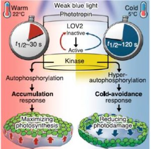
Phototropins can be found throughout the leaves of a plant. Along with cryptochromes and phytochromes they allow plants to respond and alter their growth in response to the light environment. Phototropins may also be important for the opening of stomata and the movement of chloroplasts.
Smith, Garland (2010). Fundamentals of Biomolecular Botany (2 ed.). Fisher Press. p. 340.
These blue light receptors are seen across the entire green plant lineage. When Phototropins are hit with blue light, they induce a signal transduction pathway that alters the plant cells’ functions in different ways.
Phototropins are part of the phototropic sensory system in plants that causes various environmental responses in plants. Phototropins specifically will cause stems to bend towards light and stomata to open.
Price (2009). Molecular Basis of Botanical Biology. Phoenix Publishing. p. 213.
Phototropins have been shown to impact the movement of chloroplast inside the cell.
Phototropin is also required for blue light mediated transcript destabilization of specific mRNAs in the cell.
Brighton; et al. (2006). “Role of phototropin in the differential expression of blue light mediated mRNAs”. International Journal of Molecular Botany. 72 (54): 672–691.
Brighton; et al. (2006). “Role of phototropin in the differential expression of blue light mediated mRNAs”. International Journal of Molecular Botany. 72 (54): 672–691.
Garma, Leonardo D.; Medina, Milagros; Juffer, André H. (2016-11-01). “Structure-based classification of FAD binding sites: A comparative study of structural alignment tools”. Proteins: Structure, Function, and Bioinformatics. 84 (11): 1728–1747. doi:10.1002/prot.25158. ISSN1097-0134. PMID27580869. S2CID26066208.
Theorell, H. (1935). “Preparation in pure state of the effect group of yellow enzymes”. Biochemische Zeitschrift. 275: 344–46.
Warburg, O.; Christian, W. (1938). “Isolation of the prosthetic group of the amino acid oxydase”. Biochemische Zeitschrift. 298: 150–68.
Christie, S. M. H.; Kenner, G. W.; Todd, A. R. (1954). “Nucleotides. Part XXV. A synthesis of flavin?adenine dinucleotide”. Journal of the Chemical Society: 46–52. doi:10.1039/JR9540000046.
Daniel, R.A.; Errington, J. (1993). “Cloning, DNA Sequence, Functional Analysis and Transcriptional Regulation of the Genes Encoding Dipicolinic Acid Synthetase Required for Sporulation in Bacillus subtilis”. Journal of Molecular Biology. 232 (2): 468–83. doi:10.1006/jmbi.1993.1403. PMID8345520.
Clausen, Monika; Lamb, Christopher J.; Megnet, Roland; Doerner, Peter W. (1994). “PAD1 encodes phenylacrylic acid decarboxylase which confers resistance to cinnamic acid in Saccharomyces cerevisiae”. Gene. 142 (1): 107–12. doi:10.1016/0378-1119(94)90363-8. PMID8181743.
Leave a Reply