δ-Aminolevulinic acid (also dALA, δ-ALA, 5ALA or 5-aminolevulinic acid), an endogenous non-proteinogenic amino acid, is the first compound in the porphyrin synthesis pathway, the pathway that leads to heme[Gardener LC, Cox TM (1988). “Biosynthesis of heme in immature erythroid cells”. The Journal of Biological Chemistry. 263: 6676–6682. doi:10.1016/S0021-9258(18)68695-8.] in mammals, as well as chlorophyll[Von Wettstein D, Gough S, Kannangara CG (July 1995). “Chlorophyll Biosynthesis”. The Plant Cell. 7 (7): 1039–1057. doi:10.1105/tpc.7.7.1039. PMC160907. PMID12242396.] in plants. 5ALA is used in photodynamic detection and surgery of cancer.[Wagnières, G.., Jichlinski, P., Lange, N., Kucera, P., Van den Bergh, H. (2014). Detection of Bladder Cancer by Fluorescence Cystoscopy: From Bench to Bedside – the Hexvix Story. Handbook of Photomedicine, 411-426.][Eyüpoglu IY, Buchfelder M, Savaskan NE (March 2013). “Surgical resection of malignant gliomas-role in optimizing patient outcome”. Nature Reviews. Neurology. 9 (3): 141–151. doi:10.1038/nrneurol.2012.279. PMID23358480. S2CID20352840.][Stummer W, Pichlmeier U, Meinel T, Wiestler OD, Zanella F, Reulen HJ (May 2006). “Fluorescence-guided surgery with 5-aminolevulinic acid for resection of malignant glioma: a randomised controlled multicentre phase III trial”. The Lancet. Oncology. 7 (5): 392–401. doi:10.1016/s1470-2045(06)70665-9. PMID16648043.][Eyüpoglu IY, Hore N, Savaskan NE, Grummich P, Roessler K, Buchfelder M, Ganslandt O (2012). “Improving the extent of malignant glioma resection by dual intraoperative visualization approach”. PLOS ONE. 7 (9): e44885. Bibcode:2012PLoSO…744885E.] As a precursor of a photosensitizer, 5ALA is also used as an add-on agent for photodynamic therapy.[Yew YW, Lai YC, Lim YL, Chong WS, Theng C (June 2016). “Photodynamic Therapy With Topical 5% 5-Aminolevulinic Acid for the Treatment of Truncal Acne in Asian Patients”. Journal of Drugs in Dermatology. 15 (6): 727–732. PMID27272080.] In contrast to larger photosensitizer molecules, it is predicted by computer simulations to be able to penetrate tumor cell membranes.[Erdtman E (2008). “Modelling the behavior of 5-aminolevulinic acid and its alkyl esters in a lipid bilayer”. Chemical Physics Letters. 463 (1–3): 178. Bibcode:2008CPL…463..178E. doi:10.1016/j.cplett.2008.08.021.] Photodynamic detection is the use of photosensitive drugs with a light source of the right wavelength for the detection of cancer, using fluorescence of the drug. 5ALA, or derivatives thereof, can be used to visualize bladder cancer by fluorescence imaging.[Wagnières, G.., Jichlinski, P., Lange, N., Kucera, P., Van den Bergh, H. (2014). Detection of Bladder Cancer by Fluorescence Cystoscopy: From Bench to Bedside – the Hexvix Story. Handbook of Photomedicine, 411-426.]
In non-photosynthetic eukaryotes such as animals, fungi, and protozoa, as well as the class Alphaproteobacteria of bacteria, it is produced by the enzyme ALA synthase, from glycine and succinyl-CoA. This reaction is known as the Shemin pathway, which occurs in mitochondria.[Ajioka, James; Soldati, Dominique, eds. (September 13, 2007). “22”. Toxoplasma: Molecular and Cellular Biology (1 ed.). Taylor & Francis. p. 415. ISBN9781904933342]
In plants, algae, bacteria (except for the class Alphaproteobacteria) and archaea, it is produced from glutamic acid via glutamyl-tRNA and glutamate-1-semialdehyde. The enzymes involved in this pathway are glutamyl-tRNA synthetase, glutamyl-tRNA reductase, and glutamate-1-semialdehyde 2,1-aminomutase. This pathway is known as the C5 or Beale pathway.[Beale SI (August 1990). “Biosynthesis of the Tetrapyrrole Pigment Precursor, delta-Aminolevulinic Acid, from Glutamate”. Plant Physiology. 93 (4): 1273–1279. doi:10.1104/pp.93.4.1273. PMC1062668. PMID16667613.][Willows, R.D. (2004). “Chlorophylls”. In Goodman, Robert M. Encyclopaedia of Plant and Crop Science. Marcel Dekker. pp. 258–262. ISBN0-8247-4268-0] In most plastid-containing species, glutamyl-tRNA is encoded by a plastid gene, and the transcription, as well as the following steps of C5 pathway, take place in plastids.[Biswal, Basanti; Krupinska, Karin; Biswal, Udaya, eds. (2013). Plastid Development in Leaves during Growth and Senescence (Advances in Photosynthesis and Respiration). Dordrecht: Springer. p. 508. ISBN9789400757233]
Cancer cells lack or have reduced ferrochelatase activity and this results in accumulation of Protoporphyrin IX, a fluorescent substance that can easily be visualized.[Wagnières, G.., Jichlinski, P., Lange, N., Kucera, P., Van den Bergh, H. (2014). Detection of Bladder Cancer by Fluorescence Cystoscopy: From Bench to Bedside – the Hexvix Story. Handbook of Photomedicine, 411-426.]
Lipoic acid (LA), also known as α-lipoic acid, alpha-lipoic acid (ALA) and thioctic acid, is an organosulfur compound derived from caprylic acid (octanoic acid). ALA is made in animals normally, and is essential for aerobic metabolism. It is also manufactured and is available as a dietary supplement in some countries where it is marketed as an antioxidant, and is available as a pharmaceutical drug in other countries. Lipoate is the conjugate base of lipoic acid, and the most prevalent form of LA under physiological conditions. Only the (R)-(+)-enantiomer (RLA) exists in nature and is essential for aerobic metabolism because RLA is an essential cofactor of many enzyme complexes. Lipoic acid (LA), also known as α-lipoic acid,[“Lipoic acid”. Micronutrient Information Center, Linus Pauling Institute, Oregon State University, Corvallis. 1 January 2019. Retrieved 5 November 2019][Shay, KP; Moreau, RF; Smith, EJ; Hagen, TM (June 2008). “Is alpha-lipoic acid a scavenger of reactive oxygen species in vivo? Evidence for its initiation of stress signaling pathways that promote endogenous antioxidant capacity”. IUBMB Life. 60 (6): 362–7. doi:10.1002/iub.40. PMID18409172. S2CID33008376.] alpha-lipoic acid (ALA), and thioctic acid[Reljanovic, M; Reichel, G; Rett, K; Lobisch, M; et al. (September 1999). “Treatment of diabetic polyneuropathy with the antioxidant thioctic acid (alpha-lipoic acid): A two year multicenter randomized double-blind placebo-controlled trial (ALADIN II). Alpha Lipoic Acid in Diabetic Neuropathy”. Free Radical Research. 31 (3): 171–9. doi:10.1080/10715769900300721. PMID10499773.] is an organosulfur compound derived from octanoic acid. LA contains two sulfur atoms (at C6 and C8) connected by a disulfide bond and is thus considered to be oxidized although either sulfur atom can exist in higher oxidation states.[“Lipoic acid”. Micronutrient Information Center, Linus Pauling Institute, Oregon State University, Corvallis. 1 January 2019. Retrieved 5 November 2019.] The carbon atom at C6 is chiral and the molecule exists as two enantiomers (R)-(+)-lipoic acid (RLA) and (S)-(-)-lipoic acid (SLA) and as a racemic mixture (R/S)-lipoic acid (R/S-LA). LA appears physically as a yellow solid and structurally contains a terminal carboxylic acid and a terminal dithiolane ring. For use in dietary supplement materials and compounding pharmacies, the USP established an official monograph for R/S-LA.[USP32-NF27. p. 1042.][ “Unavailable First-Time Official USP Reference Standards” (PDF). Pharmacopeial Forum. USP. 35: 26. February 2009. Archived (PDF) from the original on 5 March 2022. Retrieved 13 January 2023.]
The name is also used for the class of compounds formally derived from bilane proper by replacement of some additional hydrogen atoms by various functional groups. Natural bilanes usually have side chains substituted on the two carbons in each pyrrole ring that are not adjacent to the nitrogens. Artificial bilanes may be substituted on the bridging carbons (called meso positions).[Lindsey, J. S. (2010). “Synthetic Routes to meso-Patterned Porphyrins”. Accounts of Chemical Research. 43 (2): 300–311. doi:10.1021/ar900212t. PMID19863076.]
The parent (unsubstituted) bilane is difficult to prepare and unstable,[Claudia Ryppa, Mathias O. Senge, Sabine S. Hatscher, Erich Kleinpeter, Philipp Wacker, Uwe Schilde, and Arno Wiehe (2005): “Synthesis of Mono‐ and Disubstituted Porphyrins: A‐ and 5,10‐A2‐Type Systems”. Chemistry, A European Journal, volume 11, issue 11, pages 3427-3442. doi:10.1002/chem.20050000] but substituted derivatives are synthesized by most living organisms as intermediates in the synthesis of natural porphyrins. Substituted bilanes may also be the starting point for the synthesis of artificial porphyrins.[Lindsey, J. S. (2010). “Synthetic Routes to meso-Patterned Porphyrins”. Accounts of Chemical Research. 43 (2): 300–311. doi:10.1021/ar900212t. PMID19863076.][Claudia Ryppa, Mathias O. Senge, Sabine S. Hatscher, Erich Kleinpeter, Philipp Wacker, Uwe Schilde, and Arno Wiehe (2005): “Synthesis of Mono‐ and Disubstituted Porphyrins: A‐ and 5,10‐A2‐Type Systems”. Chemistry, A European Journal, volume 11, issue 11, pages 3427-3442. doi:10.1002/chem.20050000]
The conversion entails a reversal of the last pyrrole unit (thus swapping the acetic and propionic acid groups) and a condensation reaction that closes the macrocycle by eliminating the final hydroxyl −OH with a hydrogen atom of the first ring.
In the biosynthesis of hemes and chlorophylls, uroporphyrinogen III is converted into coproporphyrinogen III by the enzyme uroporphyrinogen III decarboxylase. In the biosynthesis of sirohemes, uroporphyrinogen III is converted by two methyl transferases to dihydrosirohydrochlorin, which is subsequently oxidized sirohydrochlorin, a precursor to the siroheme prosthetic group. If uroporphyrinogen-III synthase is not present or inactive, the hydroxymethylbilane will spontaneously cyclise into the structural isomeruroporphyrinogen I, which differs from the III isomer in that the acetic acid (“A”) and propionic acid (“P”) groups are arranged in a rotationally symmetric order, AP-AP-AP-AP. In this case, the next step produced coproporphyrinogen I, which accumulates — leading to the pathological condition congenital erythropoietic porphyria[ S. Sassa and A. Kappas (2000): “Molecular aspects of the inherited porphyrias”. Journal of Internal Medicine, volume 247, issue 2, pages 169-178. doi:10.1046/j.1365-2796.2000.00618.x]
Uroporphyrinogens are cyclic tetrapyrroles with four propionic acid groups (“P” groups) and four acetic acid groups (“A” groups). There are four forms, which vary based upon the arrangements of the “P” and “A” groups (in clockwise order): In the “I” variety (i.e. uroporphyrinogen I), the order repeats four times: AP-AP-AP-AP, In the “III” variety (i.e. uroporphyrinogen III), the fourth is reversed: AP-AP-AP-PA. This is the most common form. In the synthesis of porphyrin, it is created from the linear tetrapyrrole hydroxymethylbilane by the enzyme uroporphyrinogen III synthase, and is further converted into coproporphyrinogen III by the enzyme uroporphyrinogen III decarboxylase. The “II” and “IV” varieties can be created synthetically, but do not appear in nature.
Protoporphyrinogen IX is an organic chemical compound which is produced along the synthesis of porphyrins, a class of critical biochemicals that include hemoglobin and chlorophyll. It is a direct precursor of protoporphyrin IX. The compound is a porphyrinogen, meaning that it has a non-aromatichexahydroporphine core, which will be oxidized to a porphine core in later stages of the heme synthesis. Like most porphyrinogens, it is colorless.[citation needed] The compound is synthesized in most organisms from coproporphyrinogen III by the enzyme coproporphyrinogen oxidase. The process entails conversion of two of four propionic acid groups to vinyl groups. In coproporphyrinogen III, the substituents on the pyrrole rings have the arrangement MP-MP-MP-PM, where M and P are methyl and propionic acid, respectively. In protoporphyrinogen IX, the sequence becomes MV-MV-MP-PM, where V is vinyl. By the action of protoporphyrinogen oxidase, protoporphyrinogen IX is later converted into protoporphyrin IX, the first colored tetrapyrrole in the biosynthesis of hemes.[Paul R. Ortiz de Montellano (2008). “Hemes in Biology”. Wiley Encyclopedia of Chemical Biology. John Wiley & Sons. doi:10.1002/9780470048672.wecb221. ISBN978-0470048672.]
Protoporphyrinogen oxidase is responsible for the seventh step in biosynthesis of protoporphyrin IX. This porphyrin is the precursor to hemoglobin, the oxygen carrier in animals, and chlorophyll, the dye in plants. The enzyme catalyzes the dehydrogenation (removal of hydrogen atoms) of protoporphyrinogen IX (the product of the sixth step in the production of heme) to form protoporphyrin IX. One additional enzyme must modify protoporphyrin IX before it becomes heme. Inhibition of this enzyme is a strategy used in certain herbicides.
Clinical significance
Variegate porphyria is caused by mutations in the PPOX gene. More than 100 mutations that can cause variegate porphyria have been identified in the PPOX gene. One mutation, a substitution of the amino acidtryptophan for arginine at position 59 (also written as Arg59Trp or R59W), is found in about 95 percent of South African families with variegate porphyria. Mutations in the PPOX gene reduce the activity of the enzyme made by the gene, allowing byproducts of heme production to build up in the body. This buildup, in combination with nongenetic factors (such as certain drugs, alcohol and dieting), causes this type of porphyria.
Medlock AE, Carter M, Dailey TA, Dailey HA, Lanzilotta WN. Product release rather than chelation determines metal specificity for ferrochelatase. J Mol Biol. 2009 Oct 23;393(2):308-19. doi: 10.1016/j.jmb.2009.08.042. Epub 2009 Aug 22. PMID: 19703464; PMCID: PMC2771925.
The compound is encountered in nature in the form of complexes where the two inner hydrogen atoms are replaced by a divalent metal cation. When complexed with an iron(II) (ferrous) cation Fe2+, the molecule is called heme. Hemes are prosthetic groups in some important proteins. These heme-containing proteins include hemoglobin, myoglobin, and cytochrome c. Complexes can also be formed with other metal ions, such as zinc.[Paul R. Ortiz de Montellano (2008). “Hemes in Biology”. Wiley Encyclopedia of Chemical Biology. John Wiley & Sons. doi:10.1002/9780470048672.wecb221. ISBN978-0470048672.]
In the biosynthesis of those molecules, the metal cation is inserted into protoporphyrin IX by enzymes called chelatases. For example, ferrochelatase converts the compound into heme B (i.e. Fe-protoporphyrin IX or protoheme IX). In chlorophyll biosynthesis, the enzyme magnesium chelatase converts it into Mg-protoporphyrin IX.
Described metalloprotoporphyrin IX derivatives
Palepron is the disodium salt of protoporphyrin IX.[PubChem. “Protoporphyrin disodium”. pubchem.ncbi.nlm.nih.gov. Retrieved 2021-04-15.]
Biliverdin (from the Latin for green bile) is a green tetrapyrrolic bilepigment, and is a product of hemecatabolism.[Boron W, Boulpaep E. Medical Physiology: a cellular and molecular approach, 2005. 984-986. Elsevier Saunders, United States. ISBN1-4160-2328-3]
It is the pigment responsible for a greenish color sometimes seen in bruises.[Mosqueda, L; Burnight, K; Liao, S (2005). “The Life Cycle of Bruises in Older Adults”. Journal of the American Geriatrics Society. 53 (8): 1339–1343. doi:10.1111/j.1532-5415.2005.53406.x. PMID16078959. S2CID12394659.]
Biliverdin results from the breakdown of the heme moiety of hemoglobin in erythrocytes. Macrophages break down senescent erythrocytes and break the heme down into biliverdin along with hemosiderin, in which biliverdin normally rapidly reduces to free bilirubin.[Boron W, Boulpaep E. Medical Physiology: a cellular and molecular approach, 2005. 984-986. Elsevier Saunders, United States. ISBN1-4160-2328-3][Seyfried, H; Klicpera, M; Leithner, C; Penner, E (1976). “Bilirubin metabolism (author’s transl)”. Wiener Klinische Wochenschrift. 88 (15): 477–82. PMID793184.]
Biliverdin is seen briefly in some bruises as a green color. In bruises, its breakdown into bilirubin leads to a yellowish color.[Mosqueda, L; Burnight, K; Liao, S (2005). “The Life Cycle of Bruises in Older Adults”. Journal of the American Geriatrics Society. 53 (8): 1339–1343. doi:10.1111/j.1532-5415.2005.53406.x. PMID16078959. S2CID12394659.]
Role in disease
Biliverdin has been found in excess in the blood of humans suffering from hepatic diseases. Jaundice is caused by the accumulation of biliverdin or bilirubin (or both) in the circulatory system and tissues.[Boron W, Boulpaep E. Medical Physiology: a cellular and molecular approach, 2005. 984-986. Elsevier Saunders, United States. ISBN1-4160-2328-3] Jaundiced skin and sclera (whites of the eyes) are characteristic of liver failure.
Biliverdin is an important pigment component in avian egg shells, especially blue and green shells. Blue egg shells have a significantly higher concentration of biliverdin than brown egg shells.[Halepas, Steven; Hamchand, Randy; Lindeyer, Samuel E. D.; Brückner, Christian (2017). “Isolation of Biliverdin IXα, as its Dimethyl Ester, from Emu Eggshells”. Journal of Chemical Education. 94 (10): 1533–1537. Bibcode:2017JChEd..94.1533H. doi:10.1021/acs.jchemed.7b00449.]
Research has shown that the biliverdin of egg shells is produced from the shell gland, rather than from the breakdown of erythrocytes in the blood stream,[citation needed] although there is no evidence that the sources of the material are neither tetrapyrroles nor free haem from the blood plasma.[clarification needed][citation needed]
Along with its presence in avian egg shells, other studies have also shown that biliverdin is present in the blue-green blood of many marine fish, the blood of tobacco hornworm, the wings of moth and butterfly, the serum and eggs of frogs, and the placenta of dogs.[Fang, LS; Bada, JL (1990). “The blue-green blood plasma of marine fish”. Comparative Biochemistry and Physiology B. 97 (1): 37–45. doi:10.1016/0305-0491(90)90174-R. PMID2253479.]With dogs this can lead, in extremely rare cases, to the birth of puppies with green fur; however, the green color fades out soon after birth.[ “These Puppies Were Born with Green Fur”.] In the garfish (Belone belone) and related species, the bones are bright green because of biliverdin.[citation needed] The green coloration of many grasshoppers and lepidopteran larvae is also due to biliverdin.[Shamim, G; Ranjan, S; Pandey, D; Ramani, R (2014). “Biochemistry and biosynthesis of insect pigments” (PDF). European Journal of Entomology. 111(2): 155. doi:10.14411/eje.2014.021. Retrieved 25 June 2023.]
The main application for cyanine dyes is in biological labeling. Nevertheless, there is a wide literature on both their synthesis and uses, and cyanines are common in some CD and DVD media.
A spiropyran is a type of organic chemical compound, known for photochromic properties that provide this molecule with the ability of being used in medical and technological areas. Spiropyrans were discovered in the early twentieth century.[ Kortekaas L, Browne WR (June 2019). “The evolution of spiropyran: fundamentals and progress of an extraordinarily versatile photochrome”. Chemical Society Reviews. 48 (12): 3406–3424. doi:10.1039/C9CS00203K. PMID31150035.] However, it was in the middle twenties when Fisher and Hirshbergin observed their photochromic characteristics and reversible reaction. In 1952, Fisher and co-workers announced for the first time photochromism in spiropyrans. Since then, there have been many studies on photochromic compounds that have continued up to the present.[ Lukyanov BS, Lukyanova MB (2005). “Spiropyrans: Synthesis, Properties, and Application. A review”. Chemistry of Heterocyclic Compounds. 41 (3): 281–311. doi:10.1007/s10593-005-0148-x.][Negri RM, Prypsztejn HE (2001). “An Experiment on Photochromism and Kinetics for the Undergraduate Laboratory”. Journal of Chemical Education. 78 (5): 645. doi:10.1021/ed078p645.][Itoh K, Okamoto T, Wakita S, Niikura H, Murabayashi M (1991). “Thin films of peroxopolytungstic acids: applications to optical waveguide components”. Applied Organometallic Chemistry. 5 (4): 295. doi:10.1002/aoc.590050413.][Bertelson R (2002). Spiropyrans. Organic Photochromic and Thermochromic Compounds. Topics in Applied Chemistry. Vol. 5. pp. 11–83. doi:10.1007/0-306-46911-1_2. ISBN978-0-306-45882-8.]
As first described in 1878, the dye is prepared by the fusion of phthalic anhydride and quinaldine.
Quinaldine sulfate is an anaesthetic used in fish transportation.[Blasiola G. C. Jr. (1977). “Quinaldine sulphate, a new anaesthetic formulation for tropical marine fishes”. Journal of Fish Biology. 10 (2): 113–119(7). doi:10.1111/j.1095-8649.1977.tb04048.x.] In some Caribbean islands it is used to facilitate the collection of tropical fish from reefs.
Quinaldine is used in manufacturing anti-malaria drugs, dyes and food colorants (e.g., Quinoline Yellows, pinacyanol). It is the precursor to the pH indicator Quinaldine Red.
Quinaldine red {QR)[Salem, Ibrahim A.; El-Maazawi, M. (2001). “Kinetics and Mechanism of the Homogeneous Oxidation of Quinaldine Red by Hydrogen Peroxide”. Zeitschrift für Physikalische Chemie. 215 (5): 623–6.] is a dark green–red or black solid that does not dissolve easily in water (it is partly miscible).[Guesten, Hans (2006). “Note on the phototropic reaction of quinaldine red” (PDF). Retrieved April 12, 2013.] In addition to being used as colored indicator, quinaldine red is also used as a fluorescence probe and an agent in bleaching. Quinaldine red has the ability to fluoresce. Free quinaldine red does not fluoresce in solution when it is not bound to anything, making quinaldine red only visible by fluorescence when it is bound to something. Quinaldine red can exhibit fluorescence when it is bound to nucleic acids, which then emit radiation between 580-650 nm. [ Li, Wen-You; Miao, Kun; Wu, Hui-Ling; He, Xi-Wen; et al. (2003). “The Fluorescent Reaction Between Quinaldine Red and Nucleic Acids and its Application to Fluorescent Assay of DNA and RNA”. Microchimica Acta. 143: 33–7. doi:10.1007/s00604-003-0032-2. S2CID94415153.] The dye’s ability to bind to proteins makes it a great tag. When exposed to intensive rays such as X-rays, gamma rays, and electron beams, the dye is able to photobleach a substance. In the case of dental bleaching, a laser is the source of intensive rays. QR is dissolved in a mixture of water, ethanol, isopropyl alcohol, glycerol, and other solvents and is placed on the teeth. In the presence of oxygen, the QR and carrier particles solution uses its sensitivity to light energy to ultimately bleach teeth, making them whiter.[Kutsch, V. (2000). “Dental bleaching compositions, kits & methods”. Retrieved April 4, 2013.]
The compound exists as a mixture of two tautomers.[Horst Berneth (2008). “Methine Dyes and Pigments”. Ullmann’s Encyclopedia of Industrial Chemistry. Weinheim: Wiley-VCH. doi:10.1002/14356007.a16_487.pub2.][Han, G., Hwang, D., Lee, S. et al. Shedding new light on an old molecule: quinophthalone displays uncommon N-to-O excited state intramolecular proton transfer (ESIPT) between photobases. Sci Rep7, 3863 (2017). https://doi.org/10.1038/s41598-017-04114-9] Using other anhydrides and other quinaldine derivatives other dyes in the quinophthalone family can be prepared.
Quinoline Yellow WS is a mixture of organic compounds derived from the dye Quinoline Yellow SS (spirit soluble). Owing to the presence of sulfonate groups, the WS dyes are water-soluble (WS). It is a mixture of disulfonates (principally), monosulfonates and trisulfonates of 2-(2-quinolyl)indan-1,3-dione with a maximum absorption wavelength of 416 nm.[oint FAO/WHO Expert Committee on Food Additives (1991). Guide to specifications for general notices: general analytical techniques, identification tests, test solutions, and other reference materials (Rev. 2 [= ed. 1991] ed.). Rome: Food and Agriculture Organization of the United Nations. ISBN9789251029916.]p. 119 Quinoline Yellow is used as a greenish yellow food additive in certain countries, designated in Europe as the E number E104.[ “Current EU approved additives and their E Numbers“, Food Standards Agency website, retrieved 15 Dec 2011] In the EU and Australia, Quinoline Yellow is permitted in beverages and is used in foods, like sauces, decorations, and coatings; Quinoline Yellow is not listed as a permitted food additive in Canada or the US, where it is permitted in medicines and cosmetics and is known as D&C Yellow 10.[ Abbey J, et at. Colorants. pp 459-465 in Encyclopedia of Food Safety, Vol 2: Hazards and Diseases. Eds, Motarjemi Y et al. Academic Press, 2013. ISBN9780123786135]: 461 The Codex Alimentarius does not list it.[ Abbey J, et at. Colorants. pp 459-465 in Encyclopedia of Food Safety, Vol 2: Hazards and Diseases. Eds, Motarjemi Y et al. Academic Press, 2013. ISBN9780123786135]: 461
Health effects
Quinoline Yellow WS has not been associated with any significant long-term toxicity, is not genotoxic or carcinogenic and there is no evidence of adverse effects on reproduction or development.[ Abbey J, et at. Colorants. pp 459-465 in Encyclopedia of Food Safety, Vol 2: Hazards and Diseases. Eds, Motarjemi Y et al. Academic Press, 2013. ISBN9780123786135]Food colorants in general have been the subject of much scrutiny for their effect on health.[Amchova, Petra; Kotolova, Hana; Ruda-Kucerova, Jana “Health safety issues of synthetic food colorants” Regulatory Toxicology and Pharmacology (2015), 73(3), 914-922.doi:10.1016/j.yrtph.2015.09.026]
Cyanines were first synthesized over a century ago. They were originally used, and still are, to increase the sensitivity range of photographic emulsions, i.e., to increase the range of wavelengths which will form an image on the film, making the film panchromatic.[Berneth, Horst (2008). “Methine Dyes and Pigments”. Ullmann’s Encyclopedia of Industrial Chemistry. Weinheim: Wiley-VCH. doi:10.1002/14356007.a16_487.pub2.] Cyanines are also used in CD-R and DVD-R media. The ones used are mostly green or light blue colour, and are chemically unstable. For that reason, unstabilized cyanine discs are unsuitable for archival CD and DVD use. Recent cyanine discs contain stabilizers, typically a metal atom bonded to the cyanine molecule,[“Archival Lifespan of TDK CD-R”. cdrom2go.com. US Digital Media. Retrieved 3 April 2022.] that slow the deterioration significantly. These discs are often rated with an archival life of 75 years or more. The other dyes used in CD-Rs are phthalocyanine and azo.
Cyanine dyes are available with different modifications such as methyl, ethyl or butyl substituents, carboxyl, acetylmethoxy, and sulfo groups which alter their hydrophilicity.[CYanine dyes]
Common cyanine dyes and their uses
Because they yield brighter and more stable fluorescence, cyanines can advantageously replace conventional dyes such as fluorescein and rhodamines.
Cy3 and Cy5 are the most popular, used typically combined for 2 colors detection.
Cy3 fluoresces greenish yellow (~550 nm excitation, ~570 nm emission), while Cy5 is fluorescent in the far-red region (~650 excitation, 670 nm emission).[Jackson ImmunoResearch. “Cyanine Dyes (Cy2, Cy3, and Cy5)”. Retrieved 2008-10-31.]Cy3 can be detected by various fluorometers, imagers, and microscopes with standard filters for Tetramethylrhodamine (TRITC). Due to its high molar extinction coefficient, this dye is also easily detected by naked eye on electrophoresis gels, and in solution. Cy5 became a popular replacement for far red fluorescent dyes because of its high extinction coefficient (as small as 1 nanomol can be detected in gel electrophoresis by naked eye) and its fluorophore emission maximum in the red region, where many CCD detectors have maximum sensitivity and biological objects give low background interference.
The scanners actually use diverse laser emission wavelengths (typically 532 nm and 635 nm) and filter wavelengths (550-600 nm and 655-695 nm) to avoid background contamination. They are thus able to easily distinguish colors from Cy3 and from Cy5, and also able to quantify the amount of Cy3 and Cy5 labeling in one sample (multiparametric detection).
Other cyanine dyes are useful:
Cy3.5 can replace sulfoRhodamine 101.
Cy5.5 is a near-infrared (IR) fluorescence-emitting dye (excitation/emission maximum 678/694 nm).
Cy7 is a near-IR fluor that is invisible to the naked eye (excitation/emission maximum 750/776 nm). It is used in in vivo imaging applications, as well as the Cy7.5 dye.
Sulfo–Cyanine dyes bear one or two sulfo groups, rendering the Cy dye water-soluble, but tri- and quadri-sulfonated forms are available for even higher water solubility.[CYanine dyes]PEGylation is another modification that confers hydrophilicity, not only to the dye but also to the labeled conjugate.
Nomenclature and structure
The Cy3 and Cy5 nomenclature was first proposed by Ernst, et al.[Ernst LA, Gupta RK, Mujumdar RB, Waggoner AS (Jan 1989). “Cyanine dye labeling reagents for sulfhydryl groups”. Cytometry. 10 (1): 3–10. doi:10.1002/cyto.990100103. PMID2917472.]in 1989, and is non-standard since it gives no hint of their chemical structures. In the original paper the number designated the count of the methines (as shown), and the side chains were unspecified. Due to this ambiguity various structures are designated Cy3 and Cy5 in the literature. The R groups do not have to be identical. In the dyes as used they are short aliphatic chains one or both of which ends in a highly reactive moiety such as N-hydroxysuccinimide or maleimide.
While patent protection for the standard Cy series of dyes has lapsed, the trademarked Cy naming remains in place. Consequently, dyes that are identical to Cy dyes, but called different names, are now sold.
Applications
Cyanine dyes are used to label proteins, antibodies, peptides, nucleic acid probes, and any kind of other biomolecules to be used in a variety of fluorescence detection techniques: Flow cytometry, Microscopy (mainly Visible range, but also UV, IR), Microplate assays, Microarrays, as well as “light-up Probes,” and in vivo imaging.[Armitage, Bruce A. (27 January 2005). “Cyanine Dye–DNA Interactions: Intercalation, Groove Binding, and Aggregation”. DNA Binders and Related Subjects. Topics in Current Chemistry. Vol. 253. pp. 55–76. doi:10.1007/b100442. ISBN978-3-540-22835-6.]
Nucleic acid labeling
In microarray experimentsDNA or RNA is labeled with either Cy3 or Cy5 that has been synthesized to carry an N-hydroxysuccinimidyl ester (NHS-ester) reactive group. Since NHS-esters react readily only with aliphaticamine groups, which nucleic acids lack, nucleotides have to be modified with aminoallyl groups. This is done through incorporating aminoallyl-modified nucleotides during synthesis reactions. A good ratio is a label every 60 bases such that the labels are not too close to each other, which would result in quenching effects.
Protein labeling
For protein labeling, Cy3 and Cy5 dyes sometimes bear a succinimidyl group to react with amines, or a maleimide group to react with a sulfhydryl group of cysteine residues.
Cy5 is sensitive to its electronic environment. Changes in the conformation of the protein it is attached to will produce either enhancement or quenching of the emission. The rate of this change can be measured to determine enzyme kinetic parameters. The dyes can be used for similar purposes in FRET experiments.
Cy3 and Cy5 are used in proteomics experiments so that samples from two sources can be mixed and run together through the separation process.[19][20] This eliminates variations due to differing experimental conditions that are inevitable if the samples were run separately. These variations make it extremely difficult, if not impossible, to use computers to automate the acquisition of the data after the separation is complete. Using these dyes makes the automation trivial.
Etymology
The word cyanin is from the English word “cyan”, which conventionally means a shade of blue-green (close to “aqua”) and is derived from the Greek κυάνεος/κυανοῦς kyaneos/kyanous which means a somewhat different color: “dark blue”.
Bilirubin (BR) (from the Latin for “red bile”) is a red-orange compound that occurs in the normal catabolic pathway that breaks down heme in vertebrates. This catabolism is a necessary process in the body’s clearance of waste products that arise from the destruction of aged or abnormal red blood cells.[ Braunstein E (3 May 2019). “Overview of Hemolytic Anemia – Hematology and Oncology”. Merck Manuals Professional Edition (in Latin). Retrieved 5 May 2019.] In the first step of bilirubin synthesis, the heme molecule is stripped from the hemoglobin molecule. Heme then passes through various processes of porphyrin catabolism, which varies according to the region of the body in which the breakdown occurs. For example, the molecules excreted in the urine differ from those in the feces.[ “Bilirubin blood test”, U.S. National Library of Medicine.] The production of biliverdin from heme is the first major step in the catabolic pathway, after which the enzymebiliverdin reductase performs the second step, producing bilirubin from biliverdin.[Boron W, Boulpaep E. Medical Physiology: a cellular and molecular approach, 2005. 984–986. Elsevier Saunders, United States. ISBN1-4160-2328-3][Mosqueda L, Burnight K, Liao S (August 2005). “The life cycle of bruises in older adults”. Journal of the American Geriatrics Society. 53 (8): 1339–43. doi:10.1111/j.1532-5415.2005.53406.x. PMID16078959. S2CID12394659.]
Ultimately, bilirubin is broken down within the body, and its metabolites excreted through bile and urine; elevated levels may indicate certain diseases.[Smith ME, Morton DG (2010). “LIVER AND BILIARY SYSTEM”. The Digestive System. Elsevier. pp. 85–105. doi:10.1016/b978-0-7020-3367-4.00006-2. ISBN978-0-7020-3367-4.] It is responsible for the yellow color of healing bruises and the yellow discoloration in jaundice. Its breakdown products, such as stercobilin, cause the brown color of feces. A different breakdown product, urobilin, is the main component of the straw-yellow color in urine.[citation needed]
Strelitzia nicolai, commonly known as the wild banana or giant white bird of paradise, is a species of banana-like plants with erect woody stems reaching a height of 7–8 m (23–26 ft), and the clumps formed can spread as far as 3.5 m (11 ft).
The 1.8 m (5 ft 11 in)-long leaves are grey-green and arranged like a fan at the top of the stems, similar to Ravenala madagascariensis. The inflorescence is composed of a dark blue bract, white sepals and a bluish-purple “tongue”. The entire flower can be as much as 18 cm (7.1 in) high by 45 cm (18 in) long, and is typically held just above the point where the leaf fan emerges from the stem. Flowers are followed by triangular seed capsules.[Gartenbau-Gesellschaft., Deutsche; Petersburg., Kaiserliche Russischer Gartenbau-Verein in St.; Petersburg., Russischer Gartenbau-Verein in St.; Staaten., Verein zur Beförderung des Gartenbaues in den Königlich Preussischen (1858). “Gartenflora”. Jahrg.7 (1858).][Gibbs Russell, G. E., W. G. M. Welman, E. Retief, K. L. Immelman, G. Germishuizen, B. J. Pienaar, M. Van Wyk & A. Nicholas. 1987. List of species of southern African plants. Memoirs of the Botanical Survey of South Africa 2(1–2): 1–152(pt. 1), 1–270(pt. 2), Strelitzia nicolai]
Bilirubin consists of an open-chain tetrapyrrole. It is formed by oxidative cleavage of a porphyrin in heme, which affords biliverdin. Biliverdin is reduced to bilirubin. After conjugation with glucuronic acid, bilirubin is excreted.[citation needed]
Bilirubin is structurally similar to the pigmentphycobilin used by certain algae to capture light energy, and to the pigment phytochrome used by plants to sense light. All of these contain an open chain of four pyrrolic rings.[citation needed]
Like these other pigments, some of the double-bonds in bilirubin isomerize when exposed to light. This isomerization is relevant to the phototherapy of jaundiced newborns: the E,Z-isomers of bilirubin formed upon light exposure are more soluble than the unilluminated Z,Z-isomer, as the possibility of intramolecular hydrogen bonding is removed.[McDonagh AF, Palma LA, Lightner DA (April 1980). “Blue light and bilirubin excretion”. Science. 208 (4440): 145–51. Bibcode:1980Sci…208..145M. doi:10.1126/science.7361112. PMID7361112.] Increased solubility allows the excretion of unconjugated bilirubin in bile.
Some textbooks and research articles show the incorrect geometric isomer of bilirubin.[“Bilirubin’s Chemical Formula”. Archived from the original on 4 May 2011. Retrieved 14 August 2007.] The naturally occurring isomer is the Z,Z-isomer.
In the liver, bilirubin is conjugated with glucuronic acid by the enzyme glucuronyltransferase, first to bilirubin glucuronide and then to bilirubin diglucuronide, making it soluble in water: the conjugated version is the main form of bilirubin present in the “direct” bilirubin fraction. Much of it goes into the bile and thus out into the small intestine. Though most bile acid is reabsorbed in the terminal ileum to participate in enterohepatic circulation, conjugated bilirubin is not absorbed and instead passes into the colon.[Cheifetz AS (2010). Oxford American Handbook of Gastroenterology and Hepatology. Oxford: Oxford University Press, USA. p. 165. ISBN978-0199830121.]
There, colonic bacteria deconjugate and metabolize the bilirubin into colorless urobilinogen, which can be oxidized to form urobilin and stercobilin. Urobilin is excreted by the kidneys to give urine its yellow color and stercobilin is excreted in the feces giving stool its characteristic brown color. A trace (~1%) of the urobilinogen is reabsorbed into the enterohepatic circulation to be re-excreted in the bile.[Kuntz, Erwin (2008). Hepatology: Textbook and Atlas. Germany: Springer. p. 38. ISBN978-3-540-76838-8.]
Conjugated bilirubin’s half-life is shorter than delta bilirubin.[Sullivan KM, Gourley GR (2011). “Jaundice”. Pediatric Gastrointestinal and Liver Disease. Elsevier. pp. 176–186.e3. doi:10.1016/b978-1-4377-0774-8.10017-x. ISBN978-1-4377-0774-8.]
Delta bilirubin
Although the terms direct and indirect bilirubin are used equivalently with conjugated and unconjugated bilirubin, this is not quantitatively correct, because the direct fraction includes both conjugated bilirubin and δ bilirubin.[citation needed]
Delta bilirubin is albumin-bound conjugated bilirubin.[Tietze KJ (2012). “Review of Laboratory and Diagnostic Tests”. Clinical Skills for Pharmacists. Elsevier. pp. 86–122. doi:10.1016/b978-0-323-07738-5.10005-5. ISBN978-0-323-07738-5.] In the other words, delta bilirubin is the kind of bilirubin covalently bound to albumin, which appears in the serum when hepatic excretion of conjugated bilirubin is impaired in patients with hepatobiliary disease.[ Moyer KD, Balistreri WF (2011). “Liver Disease Associated with Systemic Disorders”. In Kliegman RM, Stanton BF, St Geme JW, Schor NF, Behrman RE (eds.). Nelson Textbook of Pediatrics. Saunders. p. 1405. ISBN978-1-4377-0755-7.] Furthermore, direct bilirubin tends to overestimate conjugated bilirubin levels due to unconjugated bilirubin that has reacted with diazosulfanilic acid, leading to increased azobilirubin levels (and increased direct bilirubin).
The half-life of delta bilirubin is equivalent to that of albumin since the former is bound to the latter, yields 2–3 weeks.[ Kalakonda A, John S (2019). “Physiology, Bilirubin article-18281”. StatPearls. Treasure Island (FL): StatPearls Publishing. PMID29261920. Retrieved 22 December 2019. This fraction of conjugated bilirubin gets covalently bound to albumin, and is called delta bilirubin or delta fraction or biliprotein. As the delta bilirubin is bound to albumin, its clearance from serum takes about 12–14 days (equivalent to the half-life of albumin) in contrast to the usual 2 to 4 hours (half-life of bilirubin).][“Unconjugated Hyperbilirubinemia: Practice Essentials, Background, Pathophysiology”. Medscape Reference. 4 March 2019. Retrieved 6 May 2019.]
A free-of-bound bilirubin has a half-life of 2 to 4 hours.[ Kalakonda A, John S (2019). “Physiology, Bilirubin article-18281”. StatPearls. Treasure Island (FL): StatPearls Publishing. PMID29261920. Retrieved 22 December 2019. This fraction of conjugated bilirubin gets covalently bound to albumin, and is called delta bilirubin or delta fraction or biliprotein. As the delta bilirubin is bound to albumin, its clearance from serum takes about 12–14 days (equivalent to the half-life of albumin) in contrast to the usual 2 to 4 hours (half-life of bilirubin).]
Under normal circumstances, only a very small amount, if any, of urobilinogen, is excreted in the urine. If the liver’s function is impaired or when biliary drainage is blocked, some of the conjugated bilirubin leaks out of the hepatocytes and appears in the urine, turning it dark amber. However, in disorders involving hemolytic anemia, an increased number of red blood cells are broken down, causing an increase in the amount of unconjugated bilirubin in the blood. Because the unconjugated bilirubin is not water-soluble, one will not see an increase in bilirubin in the urine. Because there is no problem with the liver or bile systems, this excess unconjugated bilirubin will go through all of the normal processing mechanisms that occur (e.g., conjugation, excretion in bile, metabolism to urobilinogen, reabsorption) and will show up as an increase of urobilinogen in the urine. This difference between increased urine bilirubin and increased urine urobilinogen helps to distinguish between various disorders in those systems.[ Roxe, D. M.; Walker, H. K.; Hall, W. D.; Hurst, J. W. (1990). “Urinalysis”. Clinical Methods: The History, Physical, and Laboratory Examinations. Butterworths. ISBN9780409900774. PMID21250145.]
Unbound bilirubin (Bf) levels can be used to predict the risk of neurodevelopmental handicaps within infants.[Hegyi, T.; Chefitz, D.; Weller, A.; Huber, A; Carayannopoulos, M.; Kleinfeld, A. (2020). “Unbound bilirubin measurements in term and late-preterm infants”. Journal of Maternal-Fetal & Neonatal Medicine. 35 (8): 1532–1538. doi:10.1080/14767058.2020.1761318. PMC7609464. PMID32366186.] Unconjugated hyperbilirubinemia in a newborn can lead to accumulation of bilirubin in certain brain regions (particularly the basal nuclei) with consequent irreversible damage to these areas manifesting as various neurological deficits, seizures, abnormal reflexes and eye movements. This type of neurological injury is known as kernicterus. The spectrum of clinical effect is called bilirubin encephalopathy. The neurotoxicity of neonatal hyperbilirubinemia manifests because the blood–brain barrier has yet to develop fully,[dubious – discuss] and bilirubin can freely pass into the brain interstitium, whereas more developed individuals with increased bilirubin in the blood are protected. Aside from specific chronic medical conditions that may lead to hyperbilirubinemia, neonates in general are at increased risk since they lack the intestinal bacteria that facilitate the breakdown and excretion of conjugated bilirubin in the feces (this is largely why the feces of a neonate are paler than those of an adult). Instead the conjugated bilirubin is converted back into the unconjugated form by the enzyme β-glucuronidase (in the gut, this enzyme is located in the brush border of the lining intestinal cells) and a large proportion is reabsorbed through the enterohepatic circulation. In addition, recent studies point towards high total bilirubin levels as a cause for gallstones regardless of gender or age.[Zeng, D.; Wu, H.; Huang, Q.; Zeng, A.; Yu, Z.; Zhong, Z. (2021). “High levels of serum triglyceride, low-density lipoprotein cholesterol, total bile acid, and total bilirubin are risk factors for gallstones”. Clinical Laboratory. 67 (8): 1905–1913. doi:10.7754/Clin.Lab.2021.201228. PMID34383399. S2CID234775572. Retrieved 11 November 2021 – via PubMed.]
Bilirubin is degraded by light. Blood collection tubes containing blood or (especially) serum to be used in bilirubin assays should be protected from illumination. For adults, blood is typically collected by needle from a vein in the arm. In newborns, blood is often collected from a heel stick, a technique that uses a small, sharp blade to cut the skin on the infant’s heel and collect a few drops of blood into a small tube. Non-invasive technology is available in some health care facilities that will measure bilirubin by using an instrument placed on the skin (transcutaneous bilirubin meter)[citation needed]
Bilirubin (in blood) is found in two forms:
“Conjugated bilirubin”
“Unconjugated bilirubin”
Note: Conjugated bilirubin is often incorrectly called “direct bilirubin” and unconjugated bilirubin is incorrectly called “indirect bilirubin”. Direct and indirect refer solely to how compounds are measured or detected in solution. Direct bilirubin is any form of bilirubin which is water-soluble and is available in solution to react with assay reagents; direct bilirubin is often made up largely of conjugated bilirubin, but some unconjugated bilirubin (up to 25%) can still be part of the “direct” bilirubin fraction. Likewise, not all conjugated bilirubin is readily available in solution for reaction or detection (for example, if it is hydrogen bonding with itself) and therefore would not be included in the direct bilirubin fraction.[citation needed]
Total bilirubin (TBIL) measures both BU and BC. Total bilirubin assays work by using surfactants and accelerators (like caffeine) to bring all of the different bilirubin forms into solution where they can react with assay reagents. Total and direct bilirubin levels can be measured from the blood, but indirect bilirubin is calculated from the total and direct bilirubin.
The bilirubin level found in the body reflects the balance between production and excretion. Blood test results are advised to always be interpreted using the reference range provided by the laboratory that performed the test.
Hyperbilirubinemia
Hyperbilirubinemia is a higher-than-normal level of bilirubin in the blood.
Mild rises in bilirubin may be caused by:
Hemolysis or increased breakdown of red blood cells
Gilbert’s syndrome – a genetic disorder of bilirubin metabolism that can result in mild jaundice, found in about 5% of the population
Rotor syndrome: non-itching jaundice, with rise of bilirubin in the patient’s serum, mainly of the conjugated type
Moderate[clarification needed] rise in bilirubin may be caused by:
Hemoglobin acts to transport oxygen which the body receives to all body tissue via blood vessels. Over time, when red blood cells need to be replenished, the hemoglobin is broken down in the spleen; it breaks down into two parts: heme group consisting of iron and bile and protein fraction. While protein and iron are utilized to renew red blood cells, pigments that make up the red color in blood are deposited into the bile to form bilirubin.[Point WW (April 1958). “Jaundice”. The American Journal of Nursing. 58 (4): 556–7. PMID13508735.] Jaundice leads to raised bilirubin levels that in turn negatively remove elastin-rich tissues.[Greenberg DA (December 2002). “The jaundice of the cell”. Proceedings of the National Academy of Sciences of the United States of America. 99 (25): 15837–9. Bibcode:2002PNAS…9915837G. doi:10.1073/pnas.012685199. PMC138521. PMID12461187. S2CID30298986.]Jaundice may be noticeable in the sclera of the eyes at levels of about 2 to 3 mg/dl (34 to 51 μmol/L),[Merck Manual Jaundice Last full review/revision July 2009 by Steven K. Herrine] and in the skin at higher levels.[For conversion, 1 mg/dl = 17.1 μmol/L.]
Jaundice is classified, depending upon whether the bilirubin is free or conjugated to glucuronic acid, into conjugated jaundice or unconjugated jaundice.[citation needed]
Urine tests
Urine bilirubin may also be clinically significant.MedlinePlus Encyclopedia: Bilirubin – urine</ref> Bilirubin is not normally detectable in the urine of healthy people. If the blood level of conjugated bilirubin becomes elevated, e.g. due to liver disease, excess conjugated bilirubin is excreted in the urine, indicating a pathological process.[“Urinalysis: three types of examinations”. Lab Tests Online (USA). Retrieved 16 August 2013.] Unconjugated bilirubin is not water-soluble and so is not excreted in the urine. Testing urine for both bilirubin and urobilinogen can help differentiate obstructive liver disease from other causes of jaundice.[Roxe, D. M.; Walker, H. K.; Hall, W. D.; Hurst, J. W. (1990). “Urinalysis”. Clinical Methods: The History, Physical, and Laboratory Examinations. Butterworths. ISBN9780409900774. PMID21250145.]
History
In ancient history, Hippocrates discussed bile pigments in two of the four humours in the context of a relationship between yellow and black biles.[Watson, Cecil J. (1977). “Historical Review of Bilirubin Chemistry”. In Berk, Paul D. (ed.). International Symposium on Chemistry and Physiology of Bile Pigments. U.S. Department of Health, Education, and Welfare, Public Health Service, National Institutes of Health. pp. 3–16.]
Hippocrates visited Democritus in Abdera who was regarded as the expert in melancholy “black bile”.[Watson, Cecil J. (1977). “Historical Review of Bilirubin Chemistry”. In Berk, Paul D. (ed.). International Symposium on Chemistry and Physiology of Bile Pigments. U.S. Department of Health, Education, and Welfare, Public Health Service, National Institutes of Health. pp. 3–16.]
Relevant documentation emerged in 1827 when M.Louis Jacques Thénard examined the biliary tract of an elephant that had died at a Paris zoo. He observed dilated bile ducts were full of yellow magma, which he isolated and found to be insoluble in water. Treating the yellow pigment with hydrochloric acid produced a strong green color. Thenard suspected the green pigment was caused by impurities derived from mucus of bile.[Watson, Cecil J. (1977). “Historical Review of Bilirubin Chemistry”. In Berk, Paul D. (ed.). International Symposium on Chemistry and Physiology of Bile Pigments. U.S. Department of Health, Education, and Welfare, Public Health Service, National Institutes of Health. pp. 3–16.]
Leopold Gmelin experimented with nitric acid in 1826 to establish the redox behavior in change from bilirubin to biliverdin, although the nomenclature did not exist at the time.[Watson, Cecil J. (1977). “Historical Review of Bilirubin Chemistry”. In Berk, Paul D. (ed.). International Symposium on Chemistry and Physiology of Bile Pigments. U.S. Department of Health, Education, and Welfare, Public Health Service, National Institutes of Health. pp. 3–16.]
German chemist and professor at the University of Heidelberg He worked on the red prussiate and created Gmelin’s test, and wrote his Handbook of Chemistry, which over successive editions became a standard reference work still in use.
Focus of his research was the Black pigment of oxen and calves eyes, outcome of this work was also the subject of Gmelins dissertation.
a close cooperation with Friedrich Tiedemann evolved with time. The two published “The digestion after tests” in 1826 and established the basis of the physiological chemistry. In the field of digestive chemistry Gmelin later discovered more components of bile and introduced Gmelin’s test. When Friedrich Wöhler worked on complex cyanogen compounds in 1822, Gmelin assisted him and discovered the Red prussiate.
From 1833 to 1838 Gmelin owned a paper mill in the north of Heidelberg situated Schriesheim, he had taken it over in the hope of profit. However, the work in the mill showed to be very time- and money-consuming and at the expense of his academic activity.
In 1817 the first volume of Gmelin’s Handbook of Chemistry was published. By 1843 it had grown in the fourth edition to 9 volumes. In this edition Gmelin included atomic theory and devoted much more space to the increasingly important organic chemistry. The Handbuch was published in print until the 8th edition in 1990, with an online database, which is less complete and less up-to-date than the print edition.[“LibGuides: Chemistry: Gmelin Handbook”. University of Texas. 29 July 2022.]
The terms ester and ketone (from German Aketon, meaning acetone) were introduced by Gmelin. Until his death Gmelin worked on the fifth edition of the handbook, which had become a valuable source of chemical information and documentation.
He also established the basis of an unambiguous classification of inorganic substances, later named the Gmelin system.
At the age of 60 Gmelin suffered a first stroke, and another in August 1850. In both strokes the right half of his body was affected; he was able to recover from the paralysis, but remained debilitated. In the spring of 1851 Gmelin applied for retirement, which was granted a few months later. In the two following years he suffered increasingly from the effects of a brain illness, at nearly 65 years Leopold Gmelin died in Heidelberg on 13 April 1853
In his works Leopold Gmelin dealt with physiology, mineralogy and chemistry. His experimental work was marked by his very thorough and comprehensive way of working; also some writing talent is attributed to him.
Gmelin’s first physiological work was his dissertation on the black pigment of oxen’s and calves’ eyes, whose coloring principle he tried to fathom. Despite the simplest chemical means he could describe the properties of the pigment and recognized the carbon rightly as the cause of staining. Gmelin’s most important physiological work was the 1826 released digestion by experiments, which he made together with Friedrich Tiedemann. The work, which also described many new working techniques, contained groundbreaking insights into the gastric juice, in which they found hydrochloric acid, and bile, in which Gmelin and Tiedemann among others discovered cholesterol and taurine. Introduced by Gmelin, Gmelin’s test enabled the detection of bile constituents in the urine of people suffering from jaundice. Furthermore, Gmelin and Tiedemann delivered a new, more refined view of the absorption of nutrients through the gastrointestinal tract; they were the founders of modern physiology.
The mineralogical works of Gmelin were analyses of various minerals, such as the Hauyne with which he made his habilitation in Göttingen, or the Laumontite and the Cordierite. In addition, Gmelin also analysed mineral waters and in 1825 published the work try of a new chemical mineral system, since he knew that the time’s usual division on outer or physical characteristics was inadequate. Leopold Gmelin’s mineral system was taken largely critical among experts, but the basic idea of an order based on the chemical composition proved to be useful.
Gmelin released the Handbook of theoretical chemistry, which was continued as the Gmelin Handbook of Inorganic Chemistry until 1997 in about 800 volumes by the Gmelin Institute, and it is continued by the Gesellschaft Deutscher Chemiker as a database. The manual, even during his lifetime his most important work, was initially intended to be a textbook, which should unite the whole chemical knowledge at that time. Due to the enormous increase in knowledge and the associated development of the handbook into a reference book, Gmelin published a compact textbook of chemistry in 1844. His chemical achievements include the discovery of the Croconic acid; he thus had synthesised the first cyclic organic compound, and the previously mentioned discovery of the red prussiate.
Leopold Gmelin also developed a forerunner of the Periodic table and improved chemical equipment.
Chemische Untersuchung des schwarzen Pigments der Ochsen- und Kälberaugen, nebst einigen physiologischen Bemerkungen über dasselbe, Dissertation, Göttingen 1812, in Latein. Schweiggers Journ. 10, S. 507–547, 1814
Oryktognostische und chemische Beobachtungen über den Haüyn und einige mit ihm vorkommende Fossilien, nebst geognostischen Bemerkungen über die Berge des alten Latiums, Schweiggers Journ. 15 S. 1-41, 1815; Ann. Phil. Thomson 4, S. 115-122; 193-199, 1814
Leopold Gmelin, Friedrich Wöhler: Neue Cyanverbindungen, Schweiggers Journ. 36 S. 230–235, 1822
Versuch eines neuen chemischen Mineralsystems, Taschenbuch gesammte Mineralog. 19, I S. 322-334; 418-474; 490-507, 1825, II S. 33-77; 97-148, 1825
Friedrich Tiedemann, Leopold Gmelin: Die Verdauung nach Versuchen, Heidelberg und Leipzig 1826, 2 Bde.
Lehrbuch der Chemie zum Gebrauche bei Vorlesungen auf Universitäten, in Militärschulen, polytechnischen Anstalten, Realschulen etc. sowie zum Selbstunterrichte, Heidelberg, Universitätsbuchhandlung Karl Winter, 1844
The term biliverdin was coined by Jöns Jacob Berzelius in 1840, although he preferred “bilifulvin” (yellow/red) over “bilirubin” (red).
Rudolf Virchow in 1847 recognized hematoidin to be identical to bilirubin.[Lightner DA (2013). “Early Scientific Investigations”. Bilirubin: Jekyll and Hyde Pigment of Life. Progress in the Chemistry of Organic Natural Products. Vol. 98. pp. 9–179. doi:10.1007/978-3-7091-1637-1_2. ISBN978-3-7091-1636-4.] It is not always distinguished from hematoidin, which one modern dictionary defines as synonymous with it[Merriam-Webster, Merriam-Webster’s Unabridged Dictionary, Merriam-Webster, archived from the original on 25 May 2020, retrieved 14 January 2018.] but another defines as “apparently chemically identical with bilirubin but with a different site of origin, formed locally in the tissues from hemoglobin, particularly under conditions of reduced oxygen tension.”[Elsevier, Dorland’s Illustrated Medical Dictionary, Elsevier, archived from the original on 11 January 2014, retrieved 14 January 2018.][Watson, Cecil J. (1977). “Historical Review of Bilirubin Chemistry”. In Berk, Paul D. (ed.). International Symposium on Chemistry and Physiology of Bile Pigments. U.S. Department of Health, Education, and Welfare, Public Health Service, National Institutes of Health. pp. 3–16.]
The synonymous identity of bilirubin and hematoidin was confirmed in 1923 by Fischer and Steinmetz using analytical crystallography.[Watson, Cecil J. (1977). “Historical Review of Bilirubin Chemistry”. In Berk, Paul D. (ed.). International Symposium on Chemistry and Physiology of Bile Pigments. U.S. Department of Health, Education, and Welfare, Public Health Service, National Institutes of Health. pp. 3–16.]
Origins pertaining to the physiological activity of bilirubin were described by Ernst Stadelmann in 1891, who may have observed the biotransformation of infused hemoglobin into bilirubin possibly inspired by Ivan Tarkhanov‘s 1874 works.[Watson, Cecil J. (1977). “Historical Review of Bilirubin Chemistry”. In Berk, Paul D. (ed.). International Symposium on Chemistry and Physiology of Bile Pigments. U.S. Department of Health, Education, and Welfare, Public Health Service, National Institutes of Health. pp. 3–16.]
Among his numerous contributions was the discovery of the skin galvanic reflex (1889). However, Tarkhnishvili’s most significant contribution was the discovery of the influence of X-rays on the central nervous system, animal behavior, the heart and circulation, and embryonic development (1896-1903). Indeed, these works have given rise to a new field in science as Radiobiology.
His greatest interest was in electrophysiology, which was a direct continuation of the work of I. M. Sechenov, of whom Tarkhanov was one of the first disciples. Tarkhanov engaged in experimental studies on the phenomena of summation in the nervous system (1869). He also studied the influence of compressed air, oxygen, and carbonic acid on nervous irritability (1876). He described the formation of bile pigments in animals and humans (1874) and was one of the first to show (1871) the restoration of fading functions in anemic animals by infusing saline in the body. He dominated work in the field of the physiology of aging (1891) and many other topics. Tarkhanov was one of the first to investigate hypnotic suggestion. Tarkhanov’s books, Hypnotism, Suggestion and Mind-reading (1886; translated into French in 1891) and Suggestion and Hypnotism (1905) aroused wide public interest.
In 1885 experiments on cutting and artificial emptying of the seminal vesicles, Tarkhanov showed that the latter played the crucial role in the generation of sexual excitement in frogs. Proceeding from these experimental results, Tarkhanov put forward a hypothesis that filling and evacuation of the seminal vesicles were the main biological cause which led to sexual arousal and its disappearance in mammals and humans.[Тарханов И. Р. К физиологии полового аппарата у лягушки (On Physiology of the Reproductive system in frogs). — «Русская медицина (Russian medicine)», 1885, №30–32, с. 1–26. See also in German: Tarchanoff, J. R., Arch. f. d. g. Physiol. des Mensches u. d. Thierc., 40, 330 (1887).]
Tarkhanov is probably best known as a pioneer of psychophysiology and radiobiology. In 1889, he was the first to observe and document the psychogalvanic reflex, i.e., variations in skin electrical potentials in the absence of any external stimuli. Tarkhanov’s method is still used today to measure skin potential. It records weak current actually produced by the body. Tarkhanov demonstrated that not only physical stimuli, but also mental activity, resulted in skin potential changes. The skin galvanic reflex is still used in applied psychophysiology as part of the polygraph in lie detection in which changes are recorded in several physiological variables while the subject is asked a series of questions pertaining to a specific issue under investigation.[Handbook of Clinical and Experimental Neuropsychology (eds. Gianfranco Denes, Luigi Pizzamiglio). Psychology Press, 1999. ISBN9780863775420. Page 33.]
After irradiating frogs and insects with X-rays in early 1896, several weeks after Röntgen‘s discovery, Tarkhanov concluded that these newly discovered rays not only photograph, but also “affect the living function”. These experiments signaled the birth of radiobiology.[ Y. B. Kudriashov. Radiation Biophysics. ISBN9781600212802. Page xxi.]
Tarkhanov found a marked attenuation of excitability and a total suppression of acidic reflexes. These experiments confirmed that the impairment of reflexes after X-ray exposure depended on neither analgesia nor sensitive skin but on the moderating effect of the central nervous system (CNS) itself. Studying the effects of X-rays on metabolism in the myocardium and the circulation of the heart, he concluded that all of the effects of X-rays were due to their moderating or retarding the activity of the CNS (1896). A few years later, Tarkhanov presented an extensive paper on the role of X-rays in biology and medicine (1903). Thus, his pioneer works had indeed forecast a new field of science as radiobiology.
Tarkhanov worked intensively at translating many medical and physiology textbooks, among them Technical Textbook of Histology, by L.-A. Ranvier (1876) and General Muscle and Nerve Physiology by I. Rosenthal (1879). Between the years 1892 and 1904, Tarkhanov contributed nearly 160 articles, from B to Z, in physiology and medicine to the Brockhaus and Efron Encyclopedic Dictionary. Following his resignation from the St. Petersburg Military Medical Academy, he published during the period 1897–1908 nearly 250 popular articles on a variety of topics. In these publications, Tarkhanov discussed many exciting problems of the time, such as health, hygiene, and nutrition of the people, issues of education of children and women, the organization of women’s higher medical education in Russia, and radiation safety. He appears through his writings as a progressive humanist scholar, struggling for justice in all areas of public life and many others. His great capacity for work, which could not be reflected negatively in his health, is amazing.
Ivan R. Tarkhanov played an important role in Russian and European physiology. During his research and relatively short life, he established a school of physician-investigators of various specialties. From this school emerged eminent physiologists, including V. Y. Chagovets (1873–1941), B. F. Werigo (1860–1925), V. I. Vartanov (1853–1919), N. Cybulski (1854–1919), and V. K. Anrep (1852–1927).
Tarkhanov holds a special place in the history of Georgia, Georgian culture, and education. He was the first Georgian physiologist before Ivane Beritashvili, who was himself the second outstanding Georgian physiologist from the Russian physiological school. Tarkhanov (Tarkhnishvili) was one of those bridges, through which the people of Georgia joined with the best Russian and European science and culture, in searching for more advanced education, social progress, and independence.
Tarchanoff, J.R. Über die Bildung von Gallenpigment aus Blutfarbstoff im Thierkörper. Pflüg. Arch. ges. Phys., 1874, Bd. 9, 53-65.
Tarchanoff, J.R. Du rôle des vaisseaux capillaries dans la circulation. Compt. rend. Soc. de Biol. 1875, vol. 26, T. I, Sec. 6, 331-333.
Tarchanoff, J.R. Etude sur les centre psychomoteur des animaux nouveau – nès et sur leur dèveloppments dans différentes conditions. Compt. rend. Soc. de Biol. 1878, vol. 30, 217-221.
Tarchanoff, J.R. Über die willkürliche Acceleration der Herzschläge beim Menschen. Pflüg. Arch. ges. Phys., 1885, Bd. 35, 109-137.
Tarchanoff, J.R. Zur Phisiologie des Geschlechtsapparates des Frosches. Pflüg. Arch. ges. Physiol., 1887, .Bd. 40, 330-351.
Tarchanoff, J.R. Décharges èlectrique dans la peau de l’homme sous l’influence de l’excitation des organes des sens et de differentes formes d’activitè psychique. Compt. rend. Soc. de Biol. 1889, vol. 41, 447-451.
Tarchanoff, J.R. Hypnotisme, suggestion et lecture des pensèes. (Trad. par E. Jaubert). 1-er èd., Paris, 1891; II-me èd., Paris, 1893, Masson. 164 pp.
Tarchanoff, J.R. Quelques observations sur le sommeil normal. Arch. Italiennes de Biologie, 1894, vol. 21, 318-321.
Tarchanoff, J.R. Influence de la musique sur l’homme et sur les animaux. Arch. Italiennes de Biologie, 1894, vol. 21, 313-317.
Tarkhanov, I.R. Experiments upon the action of Roentgen’s X-rays on organisms. Izvestya. St.-Peterb. Biol. Lab., 1896, no.3, 47-52. (in Russian)
Tarchanoff, J.R. Actions physiologiques des tubes de Crookes à distance. Compt. rend. Soc. de Biol. 1897, vol. 49, 740-743.
Tarchanoff, I. “Dècapitation”. Dictionnaire de Physiologie, Ch. Rischet, 1898/1899, Paris, Baillièr, 681-691.
Tarkhanov, I.R. Soul and Body. 1904, St.-Petersburg, 176 pp. (in Russian)
Tarchanoff, J.R. et Moldenhauer, T. Sur la radio-activitè induite et naturelle des plantes et sur son rôle probable dans la croissance des plantes. Bull. Int. L’acad. Sci. Cracovie. 1905, no.1, 728-734.
Cybulski N. et Tarchanoff I. A propose des poisons normaux de l’intestine. Arch. Int. Physiol., 1907, vol. 5, 257-261.
During the First World War , Barkan worked as a military doctor , later he was a department doctor in the flying school at the Lechfeld air base . From 1919 he worked as an assistant to Otto Frank in Munich and to Paul Morawitz in Würzburg . In 1923 he moved to Alexander Ellinger at Frankfurt University. From 1927 he was a private lecturer there. [The fact that he has not yet held a professorship in Frankfurt is also documented by the files in the Hessian Main State Archives, in which he was only referred to as “Dr.” and “lecturer” appears. (see web links)] In 1929 Barkan was granted leave of absence in Frankfurt [German Society for Internal Medicine: Commemorating and remembering Georg Barkan . Heuer/Wolf do not mention this leave of absence, but Benzenhöfer also counts him among those persecuted by the Frankfurt University. (Udo Benzenhöfer: “The Frankfurt University Medicine between 1933 and 1945”, p. 37). The files in the Hessian Main State Archives in Wiesbaden, especially the reparations file that is available there, also speak in favor of the continued existence of Barkan’s ongoing employment relationship in Frankfurt.] and moved to the University of Tartu in Estonia as full professor and director of the Pharmacological Institute .
In 1937 he was dismissed from the University of Tartu: The German Society for Internal Medicine states the following about the background to this : “The work as a German professor in Estonia was characterized by tensions against the background of Estonian nationalism and German-Baltic history. In 1937 Barkan was dismissed because he did not intend to learn the Estonian language.” [ German Society for Internal Medicine: Commemorating and remembering Georg Barkan]
Barkan returned to Germany via Switzerland . As a Jew, however, he no longer had any career opportunities there and emigrated in 1938 together with his wife Charlotte (nee Milch; 1894-1968) and his son Benedict Gunter (1925-2004) [ German Society for Internal Medicine: Commemorating and remembering Georg Barkan] to the USA, where he was a professor of biochemistry at Boston university .
Hessian Main State Archive Wiesbaden: Compensation procedure Georg Barkan, signature: HHStAW inventory 518 no. 46754; Files of the Hessian Ministry of Culture: case file Georg Barkan: signature: HHStAW inventory 504 no. 12040.
Plieninger and Fischer demonstrated an enzymatic oxidative loss of the alpha-methine bridge of heme resulting in a bis-lactam structure in 1942.[Watson, Cecil J. (1977). “Historical Review of Bilirubin Chemistry”. In Berk, Paul D. (ed.). International Symposium on Chemistry and Physiology of Bile Pigments. U.S. Department of Health, Education, and Welfare, Public Health Service, National Institutes of Health. pp. 3–16.]
From 1946 to 1953 he worked for Knoll AG in Ludwigshafen, where he worked on the synthesis of amino acids.
In 1953 he habilitated with his already published work at the TH Darmstadt. For formal reasons, he was not able to habilitate in Heidelberg, since Karl Freudenberg , his father-in-law, was the director of the institute there.
He and his wife were involved in environmental protection and peace politics. In 1982, for example, he sent all 519 members of the Bundestag the book The Fate of the Earth by Jonathan Schell against nuclear armament at his own expense (around 10,000 DM at the time) . He had a farm in Tuscany where he practiced organic farming.
From 1921 until his death, he held the position of Professor of Organic Chemistry at the Technical University of Munich.
Fischer’s scientific work was mostly concerned with the investigation of the pigments in blood, bile, and also chlorophyll in leaves, as well as with the chemistry of pyrrole from which these pigments are derived. Of special importance was his synthesis of bilirubin and haemin.
He received many honors for this work, and received the Nobel Prize in 1930.
Hans Fisher had mapped the composition of a hem group. In 1929 Fischer succeeded in producing the substance and proving that its ring has a central atom of iron, as he also continued studying other pigmented substances of a biological importance of biochemistry such as chlorophyll, the color that plays part in a plants photosynthesis.
Fischer also unraveled the bile pigments biliverdin (which causes the yellowish color characteristic of bruised skin) and bilirubin (which yellows skin in jaundice cases), and synthesized them in 1942 and 1944, successively.
He conducted microanalyses of more than 60,000 chemical substance, and had won the Nobel Prize for Chemistry in 1930.
The person that sparked Fischer’s interest was von Muller, his former professor and supervisor whom interests were in pyrrole pigments by inviting Fischer to work with him at the well-known Second Medical Clinic in Munich in 1910. Under Muller, he began to examine the composition of the bile pigment bilirubin, something he would continue to be engaged in during the decades that followed. Fischer’s succession also came with difficulty as many of his experiments seemed to have failed but with time Fischer was able to perfect his acknowledgement with is failed attempts.
Fischer married Wiltrud Haufe around his 50’s in the year 1935. Fischer was a man whom dedicated almost “exclusively” to his work. He continued his scientific research during Germany’s Nazi era, and committed suicide on Easter Sunday in 1945, after his laboratory and life’s work had been destroyed by the bombing in the last days of World War II.
Bickel, M H (2001), “[Henry E. Sigerist and Hans Fischer as pioneers of a medical history institute in Zurich]”, Gesnerus, vol. 58, no. 3–4, pp. 215–9, PMID11810971
Kämmerer, H (1961), “Hans Fischer (1881–1945). A reminiscence on the 80th anniversary of his birth”, Münchener Medizinische Wochenschrift (1950) (published Nov 3, 1961), vol. 103, pp. 2164–6, PMID14036988
Hans Fischer on Nobelprize.org including the Nobel Lecture, December 11, 1930
It is widely accepted that Irving London was the first to demonstrate endogenous formation of bilirubin from hemoglobin in 1950,[ “Bilirubin”. American Chemical Society. Retrieved 28 May 2021.]
London graduated from Harvard College in 1939 summa cum laude. He was on a student committee at Harvard that gave 14 refugee students the opportunity to leave Nazi-occupied Europe to study in Boston.[“School Marks Pioneer’s Passing”. hms.harvard.edu. Retrieved 2021-05-28.] London also earned a second undergraduate degree from Hebrew College in Roxbury at the same time. London delivered the graduating address at Harvard, the content of which was inspired by his thesis “The Jeffersonian Tradition in American Nationalism”. London gave serious though to attending law school after graduation, but ultimately chose to enroll in medical school.[“Irving M. London”. meded.hms.harvard.edu. Retrieved 2021-05-29.]
Welch Fellowship in Internal Medicine of the National Academy of Sciences 1949-1952
Theobald Smith Award in Medical Sciences of the American Association for the Advancement of Science 1953
Commonwealth Fund Fellowship at Institut Pasteur 1962-1963
election to American Academy of Arts and Sciences 1963
charter member in the Institute of Medicine of the National Academy of Sciences in 1970
elected member National Academy of Science 1971
board of directors for Biosciences Advisory Committee for Johnson & Johnson 1982-2003
establishment of The Irving M. London Society (HST) at Harvard Medical School[7]
The Dr. Irving M. London Teaching Award, initiated in 1986[“Community Awards”. Harvard-MIT Health Sciences and Technology. 2019-12-19. Retrieved 2021-05-29.]
Sjöstrand later demonstrated CO production from hemoglobin decomposition in 1952.[Hopper CP, Zambrana PN, Goebel U, Wollborn J (June 2021). “A brief history of carbon monoxide and its therapeutic origins”. Nitric Oxide. 111–112: 45–63. doi:10.1016/j.niox.2021.04.001. ISSN1089-8603. PMID33838343. S2CID233205099.]
14C labeled protoporphyrin biotransformation to bilirubin evidence emerged in 1966 by Cecil Watson.[Watson, Cecil J. (1977). “Historical Review of Bilirubin Chemistry”. In Berk, Paul D. (ed.). International Symposium on Chemistry and Physiology of Bile Pigments. U.S. Department of Health, Education, and Welfare, Public Health Service, National Institutes of Health. pp. 3–16.]
Upon his return to the United States in 1932, Watson began working at Minneapolis General Hospital. By 1934, Watson was assistant professor of medicine at UM. Watson lead the medical school as chairman from 1943 to 1966, stepping down for a position at Northwestern Hospital.[Schmid, Rudi. “Cecil James Watson 1901—1983” (PDF). Biographical Memoirs of the National Academy of Sciences.]
Watson was named a member of the National Academy of Sciences in 1959,[“Cecil J. Watson”. National Academy of Sciences. Retrieved October 15, 2018.] and the Cecil J. Watson Award was inaugurated in his honor by the Minneapolis Society of Internal Medicine in 1961.[“Cecil J. Watson Award”. University of Minnesota. Retrieved October 15, 2018.]
HMOX1 was first characterized by Tenhunen and Rudi Schmid upon demonstrating it as the enzyme responsible for catalyzing biotransformation of heme to bilirubin.[Hopper CP, Zambrana PN, Goebel U, Wollborn J (June 2021). “A brief history of carbon monoxide and its therapeutic origins”. Nitric Oxide. 111–112: 45–63. doi:10.1016/j.niox.2021.04.001. ISSN1089-8603. PMID33838343. S2CID233205099.]
Several labs attempted to explain the biotransformation of heme to biliverdin such as Nakajima et al. in 1962 who characterized a soluble “heme α-methenyl oxygenase”, however the findings could not be reproduced and alternative non-enzymatic explanations for their observation emerged. The earliest evidence of oxidative enzymatic biotransformation of heme to a bilin was demonstrated by Hans Plieninger and Hans Fischer in 1942.[Watson C (1977). “Historical Review of Bilirubin Chemistry”. In Berk P (ed.). Chemistry and Physiology of Bile Pigments. p. 5.] The discovery of HMOX is a case of academic linage as Fischer was the academic adviser for Cecil Watson, and Watson was an adviser for Rudi Schmid.
CO was detected in exhaled breath 1869. Felix Hoppe-Seyler developed the first qualitative carboxyhemoglobin test, and Josef von Fodor developed the first quantitative analytical test for carboxyhemoglobin. The first reported detection of naturally occurring CO in human blood occurred in 1923 by Royd Ray Sayers et al. although they discarded their data as random error.[Hopper CP, Zambrana PN, Goebel U, Wollborn J (June 2021). “A brief history of carbon monoxide and its therapeutic origins”. Nitric Oxide. 111–112: 45–63. doi:10.1016/j.niox.2021.04.001. ISSN1089-8603. PMID33838343. S2CID233205099.]Alexander Gettler confirmed CO to have a normal presence in blood in 1933, however, he attributed the finding to inevitable pollution exposure or perhaps derived from the human microbiome.[Hopper CP, De La Cruz LK, Lyles KV, Wareham LK, Gilbert JA, Eichenbaum Z, et al. (December 2020). “Role of Carbon Monoxide in Host-Gut Microbiome Communication”. Chemical Reviews. 120 (24): 13273–13311. doi:10.1021/acs.chemrev.0c00586. PMID33089988. S2CID224824871.] Sjöstrand later demonstrated CO production from hemoglobin decomposition in 1952.[Hopper CP, Zambrana PN, Goebel U, Wollborn J (June 2021). “A brief history of carbon monoxide and its therapeutic origins”. Nitric Oxide. 111–112: 45–63. doi:10.1016/j.niox.2021.04.001. ISSN1089-8603. PMID33838343. S2CID233205099.]
Earlier in 1963, Nakajima described a soluble “heme alpha-methnyl oxygeanse” which what later determined to be a non-enzymatic pathway, such as formation of a 1,2-Dioxetane intermediate at the methine bridge resulting in carbon monoxide release and biliverdin formation.[Berk, Paul D.; Berlin, Nathaniel I. (1977). International Symposium on Chemistry and Physiology of Bile Pigments. U.S. Department of Health, Education, and Welfare, Public Health Service, National Institutes of Health. pp. 27, 50.]
The drug causes hepatocellular injury, generally defined as an elevated ALT or AST by 3-fold or greater above the upper limit of normal. Often with aminotransferases much greater (5-10x) the upper limit of normal.
Among subjects showing such aminotransferase elevations, they also have elevation of their serum total bilirubin of greater than 2× the upper limit of normal, without findings of cholestasis (defined as serum alkaline phosphatase activity less than 2× the upper limit of normal).
No other reason can be found to explain the combination of increased aminotransferase and serum total bilirubin, such as viral hepatitis, alcohol abuse, ischemia, preexisting liver disease, or another drug capable of causing the observed injury.[Reuben, Adrian (March 26, 2008). “Hy’s Law Explained” (PDF). fda.gov. Retrieved December 7, 2016.]
Shapiro, Steven M. (January 2005). “Definition of the Clinical Spectrum of Kernicterus and Bilirubin-Induced Neurologic Dysfunction (BIND)”. Journal of Perinatology. 25 (1): 54–59. doi:10.1038/sj.jp.7211157. PMID15578034. S2CID19663259.
Smith, Margaret E.; Morton, Dion G. (2010). “Liver and Biliary System”. The Digestive System. Elsevier. pp. 85–105. doi:10.1016/b978-0-7020-3367-4.00006-2. ISBN978-0-7020-3367-4. However, when jaundice is present it is likely that many other potentially toxic materials have also accumulated in the blood as a consequence of their reflux from the bile or impaired secretion from the hepatocyte. This can lead to impaired mental function and malaise.
Watchko, Jon F.; Tiribelli, Claudio (2013-11-21). Ingelfinger, Julie R. (ed.). “Bilirubin-Induced Neurologic Damage — Mechanisms and Management Approaches”. The New England Journal of Medicine. 369 (21): 2021–2030. doi:10.1056/nejmra1308124. ISSN0028-4793. PMID24256380.
In Dubin–Johnson syndrome, impaired biliary excretion of bilirubin glucuronide is due to a mutation in the canalicular multiple drug-resistance protein 2 (MRP2). A darkly pigmented liver is due to polymerized epinephrine metabolites, not bilirubin.[Kumar, Vinay (2007). Robbins Basic Pathology. Elsevier. p. 639.]
Liver failure or hepatitis
If it is the liver that cannot effectively transfer the indirect bilirubin into bilirubin glucuronide and further into bilirubin di-glucuronide, the consequence will be hyperbilirubinemia or intrahepatic (or hepatocellular) jaundice. Moreover, the unconjugated hyperbilirubinemia arises in case the components of liver transfer the indirect bilirubin into bilirubin glucuronide in the rate slower than they should be.[Smith, Margaret E.; Morton, Dion G. (2010). “Liver and Biliary System”. The Digestive System. Elsevier. pp. 85–105. doi:10.1016/b978-0-7020-3367-4.00006-2. ISBN978-0-7020-3367-4.] This condition is associated with either decreased uptake of bilirubin into hepatocytes (Rotor syndrome[van de Steeg, Evita; Stránecký, Viktor; Hartmannová, Hana; Nosková, Lenka; Hřebíček, Martin; Wagenaar, Els; van Esch, Anita; de Waart, Dirk R.; Oude Elferink, Ronald P.J.; Kenworthy, Kathryn E.; Sticová, Eva; al-Edreesi, Mohammad; Knisely, A.S.; Kmoch, Stanislav; Jirsa, Milan; Schinkel, Alfred H. (2012-02-01). “Complete OATP1B1 and OATP1B3 deficiency causes human Rotor syndrome by interrupting conjugated bilirubin reuptake into the liver”. The Journal of Clinical Investigation. 122 (2): 519–528. doi:10.1172/jci59526. ISSN0021-9738. PMC3266790. PMID22232210.]) or defective intracellular protein binding. In similar fashion, the conjugated hyperbilirubinemia emerges in case the components of the liver have difficulty turning bilirubin glucuronide into bilirubin di-glucuronide.[Smith, Margaret E.; Morton, Dion G. (2010). “Liver and Biliary System”. The Digestive System. Elsevier. pp. 85–105. doi:10.1016/b978-0-7020-3367-4.00006-2. ISBN978-0-7020-3367-4.] Note that biliary duct blockage can also lead to conjugated hyperbilirubinemia but the pathophysiology is that backflow of bilirubin di-glucuronide with little indirect bilirubin and bilirubin glucuronide from bile duct through liver into blood plasma.[Smith, Margaret E.; Morton, Dion G. (2010). “Liver and Biliary System”. The Digestive System. Elsevier. pp. 85–105. doi:10.1016/b978-0-7020-3367-4.00006-2. ISBN978-0-7020-3367-4.][Weiss, Janet S.; Gautam, Anil; Lauff, John J.; Sundberg, Michael W.; Jatlow, Peter; Boyer, James L.; Seligson, David (1983-07-21). “The Clinical Importance of a Protein-Bound Fraction of Serum Bilirubin in Patients with Hyperbilirubinemia”. The New England Journal of Medicine. 309 (3): 147–150. doi:10.1056/nejm198307213090305. ISSN0028-4793. PMID6866015.] These conditions are associated with either defective intracellular protein binding (for the second time) or disturbed secretion into the bile canaliculi (Dubin-Johnson syndrome[van de Steeg, Evita; Stránecký, Viktor; Hartmannová, Hana; Nosková, Lenka; Hřebíček, Martin; Wagenaar, Els; van Esch, Anita; de Waart, Dirk R.; Oude Elferink, Ronald P.J.; Kenworthy, Kathryn E.; Sticová, Eva; al-Edreesi, Mohammad; Knisely, A.S.; Kmoch, Stanislav; Jirsa, Milan; Schinkel, Alfred H. (2012-02-01). “Complete OATP1B1 and OATP1B3 deficiency causes human Rotor syndrome by interrupting conjugated bilirubin reuptake into the liver”. The Journal of Clinical Investigation. 122 (2): 519–528. doi:10.1172/jci59526. ISSN0021-9738. PMC3266790. PMID22232210.]). Liver failure and hepatitis are the most etiological in liver-genesis hyperbilirubinemia. In case of hyperbilirubinemia due to intrahepatic or extrahepatic bile ducts blockage, e.g. gallstone, the name is given as Post-hepatic (or obstructive) jaundice.[Smith, Margaret E.; Morton, Dion G. (2010). “Liver and Biliary System”. The Digestive System. Elsevier. pp. 85–105. doi:10.1016/b978-0-7020-3367-4.00006-2. ISBN978-0-7020-3367-4.] Bilirubin concentration is not a sensitive early indicator of liver diseases as the liver may have reserved its capacity in removal of bilirubin to save energy and unreserved the previously reserved capacity when encountering a sudden rise of unconjugated bilirubin. In short, there is still a chance for an ill liver to get rid of excessive unconjugated bilirubin in the blood plasma, displaying a total bilirubin level that is within normal reference range.[Raymond, GD; Galambos, JT (1971). “Hepatic storage and excretion of bilirubin in man”. The American Journal of Gastroenterology. 55 (2): 135–44. ISSN0002-9270. PMID5580257.]
In jaundice owing to hemolysis (Prehepatic (or hemolytic) jaundice), the pathophysiology is that overproduction of bilirubin from the extravascular or intravascular hemolysis overwhelms the capacity of the liver to excrete it. The bilirubin present in the plasma is largely unconjugated in this setting as they haven’t been taken up and conjugated by the liver.[Smith, Margaret E.; Morton, Dion G. (2010). “Liver and Biliary System”. The Digestive System. Elsevier. pp. 85–105. doi:10.1016/b978-0-7020-3367-4.00006-2. ISBN978-0-7020-3367-4.] In this case, total serum bilirubin increases while the ratio of direct bilirubin to indirect bilirubin remains 96 to 4 as up to 96%-99% of bilirubin in the bile are conjugated mentioned above.[Namita Roy-Chowdhury; Jayanta Roy-Chowdhury. Sanjiv Chopra; Elizabeth B Rand; Shilpa Grover (eds.). “Bilirubin metabolism”. UpToDate. Archived from the original on 2017-10-24. Retrieved 2019-05-05.][Divers, Thomas J.; Barton, Michelle Henry (2018). “Disorders of the Liver”. Equine Internal Medicine. Elsevier. pp. 843–887. doi:10.1016/b978-0-323-44329-6.00013-9. ISBN978-0-323-44329-6. then convert biliverdin to bilirubin and release it from the cell as free, insoluble bilirubin. This form of bilirubin also is referred to as indirect-reacting or unconjugated bilirubin.]
Shapiro, Steven M. (January 2005). “Definition of the Clinical Spectrum of Kernicterus and Bilirubin-Induced Neurologic Dysfunction (BIND)”. Journal of Perinatology. 25 (1): 54–59. doi:10.1038/sj.jp.7211157. PMID15578034. S2CID19663259.
Smith, Margaret E.; Morton, Dion G. (2010). “Liver and Biliary System”. The Digestive System. Elsevier. pp. 85–105. doi:10.1016/b978-0-7020-3367-4.00006-2. ISBN978-0-7020-3367-4. However, when jaundice is present it is likely that many other potentially toxic materials have also accumulated in the blood as a consequence of their reflux from the bile or impaired secretion from the hepatocyte. This can lead to impaired mental function and malaise.
Watchko, Jon F.; Tiribelli, Claudio (2013-11-21). Ingelfinger, Julie R. (ed.). “Bilirubin-Induced Neurologic Damage — Mechanisms and Management Approaches”. The New England Journal of Medicine. 369 (21): 2021–2030. doi:10.1056/nejmra1308124. ISSN0028-4793. PMID24256380.
Bilirubin di-glucuronide is a conjugated form of bilirubin formed in bilirubin metabolism.[Chowdhury, J. R.; Chowdhury, N. R.; Wu, G.; Shouval, R.; Arias, I. M. (1981). “Bilirubin mono- and diglucuronide formation by human liver in vitro: Assay by high-pressure liquid chromatography”. Hepatology. 1 (6): 622–7. doi:10.1002/hep.1840010610. PMID6796486.] The hydrophilic character of bilirubin diglucuronide enables it to be water-soluble. It is pumped across the hepatic canalicular membrane into the bile by the transporter MRP2.[Lengyel, G.; et al. (2007-08-29). “Modulation of sinusoidal and canalicular elimination of bilirubin-glucuronides by rifampicin and other cholestatic drugs in a sandwich culture of rat hepatocytes”. Hepatology Research. Wiley. 38 (3): 300–309. doi:10.1111/j.1872-034X.2007.00255.x. PMID17760873.]
Stercobilinogen (fecal urobilinogen) is a chemical created by bacteria in the gut. It is made of broken-down hemoglobin. It is further processed to become the chemical that gives feces its brown color.[Stercobilinogen, drugs.com]Bilirubin is a pigment that results from the breakdown of the heme portion of hemoglobin. The liver conjugates bilirubin, making it water-soluble; and the conjugated form is then excreted in urine as urobilinogen. Urobilinogen is colourless and is further oxidised to stercobilin which imparts colour to feces. Darkening of feces upon standing in air is due to the oxidation of residual urobilinogens to urobilins. In the intestine, bilirubin is converted by bacteria to stercobilinogen. Stercobilinogen is absorbed and excreted by either the liver or the kidney. Stercobilinogen is oxidized to stercobilin, which is responsible for the pigmentation of feces.
In obstructive jaundice, no bilirubin reaches the small intestine, meaning that there is no formation of stercobilinogen. The lack of stercobilin and other bile pigments causes feces to become clay-colored.[Seyfried H, Klicpera M, Leithner C, Penner E (1976). “Bilirubin metabolism”. Wiener Klinische Wochenschrift. 88:477-82. PMID793184]
A 1996 study by McPhee et al. suggested that stercobilin and other related pyrrolic pigments — including urobilin, biliverdin, and xanthobilirubic acid — has potential to function as a new class of HIV-1 protease inhibitors when delivered at low micromolar concentrations. These pigments were selected due to a similarity in shape to the successful HIV-1 protease inhibitor Merck L-700,417 (N,N-Bis(2-hydroxy-1-indanyl)-2,6-diphenylmethyl-4-hydroxy-1,7-heptandiamide). Further research is suggested to study the pharmacological efficacy of these pigments.[McPhee, Fiona; Caldera, Patricia S.; Bemis, Guy W.; McDonagh, Antony F.; Kuntz, Irwin D.; Craik, Charles S. (1 December 1996). “Bile pigments as HIV-1 protease inhibitors and their effects on HIV-1 viral maturation and infectivity in vitro”. Biochemical Journal. 320 (2): 681–686. doi:10.1042/bj3200681. PMC1217983. PMID8973584.]
Urobilinogen is a colorless by-product of bilirubin reduction. It is formed in the intestines by bacterial action on bilirubin. About half of the urobilinogen formed is reabsorbed and taken up via the portal vein to the liver, enters circulation and is excreted by the kidney.
Increased amounts of bilirubin are formed in hemolysis, which generates increased urobilinogen in the gut. In liver disease (such as hepatitis), the intrahepatic urobilinogen cycle is inhibited also increasing urobilinogen levels. Urobilinogen is converted to the yellow pigmented urobilin apparent in urine.
The urobilinogen in the intestine is directly reduced to brownish colour stercobilin, which gives the feces their characteristic color. It can also be reduced to stercobilinogen, which can then be further oxidized to stercobilin.
In biliary obstruction, below-normal amounts of conjugated bilirubin reach the intestine for conversion to urobilinogen. With limited urobilinogen available for reabsorption and excretion, the amount of urobilin found in the urine is low. High amounts of the soluble conjugated bilirubin enter the circulation where they are excreted via the kidneys. These mechanisms are responsible for the dark urine and pale stools observed in biliary obstruction.
Low urine urobilinogen may result from complete obstructive jaundice or treatment with broad-spectrum antibiotics, which destroy the intestinal bacterial flora. (Obstruction of bilirubin passage into the gut or failure of urobilinogen production in the gut.)
Low urine urobilinogen levels may result from congenital enzymatic jaundice (hyperbilirubinemia syndromes) or from treatment with drugs that acidify urine, such as ammonium chloride or ascorbic acid.
Elevated levels may indicate hemolytic anaemia (excessive breakdown of red blood cells RBC), overburdening of the liver, increased urobilinogen production, re-absorption – a large hematoma, restricted liver function, hepatic infection, poisoning or liver cirrhosis.[“Urobilinogen”. Family Health Information. Retrieved 2008-03-30.][“Urobilinogen in urine”. Home test kist. Retrieved 2008-03-30.]
Nomenclature
Urobilinogen (a.k.a. D-urobilinogen) is closely related to two other compounds: mesobilirubinogen (a.k.a. I-urobilinogen) and stercobilinogen (a.k.a. L-urobilinogen). Specifically, urobilinogen can be reduced to form mesobilirubinogen, and mesobilirubinogen can be further reduced to form stercobilinogen.[ “Biochemical Pathways Map No. L5 L6”. ExPASy Bioinformatics Resource Portal. Retrieved 12 December 2011.] Confusingly, however, all three of these compounds are frequently collectively referred to as “urobilinogens”.[Henry’s clinical diagnosis and management by laboratory methods (PDF) (22nd ed.). Philadelphia, PA: Elsevier/Saunders. 2011. p. 543. ISBN978-1-4377-0974-2.]
Urobilin or urochrome is the chemical primarily responsible for the yellow color of urine. It is a linear tetrapyrrole compound that, along with the related colorless compound urobilinogen, are degradation products of the cyclic tetrapyrrole heme. Urobilin is generated from the degradation of heme, which is first degraded through biliverdin to bilirubin. Bilirubin is then excreted as bile, which is further degraded by microbes present in the large intestine to urobilinogen. Some of this remains in the large intestine, and its conversion to stercobilin gives feces their brown color. Some is reabsorbed into the bloodstream and then delivered to kidney. When urobilinogen is exposed to air, it is oxidized to urobilin, giving urine its yellow color.[ John E. Hall (2016). “The liver as an organ”. Guyton and Hall Textbook of Medical Physiology, 13th edition. Elsevier. p. 885. ISBN978-1455770052.]
Importance
Many urine tests (urinalysis) monitor the amount of urobilin in urine, as its levels can give insight on the effectiveness of urinary tract function. Normally, urine would appear as either light yellow or colorless. A lack of water intake, for example following sleep or dehydration, reduces the water content of urine, thereby concentrating urobilin and producing a darker color of urine. Obstructive jaundice reduces biliary bilirubin excretion, which is then excreted directly from the blood stream into the urine, giving a dark-colored urine but with a paradoxically low urobilin concentration, no urobilinogen, and usually with correspondingly pale faeces. Darker urine can also be due to other chemicals, such as various ingested dietary components or drugs, porphyrins in patients with porphyria, and homogentisate in patients with alkaptonuria.
Voet and Voet Biochemistry Ed 3 page 1022
Nelson, L., David, Cox M.M., .2005. “Chapter 22- Biosynthesis of Amino Acids, Nucleotides, and Related Molecules”, pp. 856, In Lehninger Principles of Biochemistry. Freeman, New York. pp. 856
Bishop, Michael, Duben-Engelkirk, Janet L., and Fody, Edward P. “Chapter 19, Liver Function, Clinical Chemistry Principles, Procedures, Correlations, 2nd Ed.” Philadelphia: copyright 1992 J.B. Lippincott Company.
Munson-Ringsrud, Karen and Jorgenson-Linné, Jean “Urinalysis and Body Fluids, A ColorText and Atlas.” St. Louis: copyright 1995 Mosby
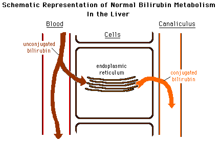
Leave a Reply