Kynureninase or L-Kynurenine hydrolase (KYNU) (EC 3.7.1.3) is a PLP dependent enzyme that catalyses the cleavage of kynurenine (Kyn) into anthranilic acid (Ant). It can also act on 3-hydroxykynurenine (to produce 3-hydroxyanthranilate) and some other (3-arylcarbonyl)-alanines.
Note: 3-Hydroxykynurenine is a metabolite of tryptophan, which filters UV light in the human lens. It is one of two pigments identified as responsible for the goldenrod crab spider‘s (Misumena vatia) yellow coloration.
- Malina, HZ; Martin, XD (1995). “Deamination of 3-hydroxykynurenine in bovine lenses: a possible mechanism of cataract formation in general”. Graefes Arch Clin Exp Ophthalmol. 233 (1): 38–44. doi:10.1007/bf00177784. PMID 7721122. S2CID 25414197.
- Schwarcz, Robert; John P. Bruno; Paul J. Muchowski; Hui-Qiu Wu (July 2012). “Kynurenines in the Mammalian Brain: When Physiology Meets Pathology”. Nature Reviews Neuroscience. 13 (7): 465–477. doi:
3-Hydroxyanthranilic acid is an intermediate in the metabolism of tryptophan. It is new antioxidant isolated from methanol extract of tempeh. It is effective in preventing autoxidation of soybean oil and powder, while antioxidant 6,7,4′-trihydroxyisoflavone is not. Tempeh or tempe is a traditional Indonesian food made from fermented soybeans. It is made by a natural culturing and controlled fermentation process that binds soybeans into a cake form. A fungus, Rhizopus oligosporus or Rhizopus oryzae, is used in the fermentation process and is also known as tempeh starter.
- Darlington LG, Forrest CM, Mackay GM, Smith RA, Smith AJ, Stoy N, Stone TW. On the Biological Importance of the 3-hydroxyanthranilic Acid: Anthranilic Acid Ratio. Int J Tryptophan Res. 2010;3:51-9. doi: 10.4137/ijtr.s4282. Epub 2010 Jun 10. PMID: 22084587; PMCID: PMC3195249.
- “3-Hydroxyanthranilic acid”. Santa Cruz Biotechnology, Inc. Retrieved 2015-09-24.
- Armarego, Wilfred L.F.; Chai, Christina L.L. (2009). Purification of Laboratory Chemicals (6th ed.). Elsevier Inc. p. 297. ISBN 978-1-85617-567-8.
- Moline, Sheldon W.; Walker, H.C.; Schweigert, B.S. (1958). “3-Hydroxyanthranilic Acid Metabolism: VII. Mechanism of Formation of Quinolinic Acid”. Journal of Biological Chemistry. 234 (4): 880–883. doi:10.1016/S0021-9258(18)70194-4. PMID 13654282. Retrieved 2015-09-24.
- Esaki, Hideo; Onozaki, Hiromichi; Kawakishi, Shunro; Osawa, Toshihiko (1996). “New Antioxidant Isolated from Tempeh”. Journal of Agricultural and Food Chemistry. 44 (3): 696. doi:10.1021/jf950454t.
- Shurtleff, William; Aoyagi, Akiko. “History of Tempeh”. www.soyinfocenter.com. p. 1. Retrieved 2018-09-16.
- “Tempeh”. Dictionary.com.
Humans express one kynureninase enzyme that is encoded by the KYNU gene located on chromosome 2.
- Alberati-Giani D, Buchli R, Malherbe P, Broger C, Lang G, Köhler C, Lahm HW, Cesura AM (July 1996). “Isolation and expression of a cDNA clone encoding human kynureninase”. Eur. J. Biochem. 239 (2): 460–8. doi:10.1111/j.1432-1033.1996.0460u.x. PMID 8706755.
- Toma S, Nakamura M, Toné S, Okuno E, Kido R, Breton J, Avanzi N, Cozzi L, Speciale C, Mostardini M, Gatti S, Benatti L (May 1997). “Cloning and recombinant expression of rat and human kynureninase”. FEBS Lett. 408 (1): 5–10. doi:10.1016/S0014-5793(97)00374-8. PMID 9180257. S2CID 36265922.
KYNU is part of the pathway for the catabolism of Trp and the biosynthesis of NAD cofactors from tryptophan (Trp).
Kynureninase catalyzes the following reaction:
L–kynurenine + H2O ↔ anthranilate + L–alanine
Structure
Kynureninase belongs to the class V group of aspartate aminotransferase superfamily of structurally homologous pyridoxal 5′-phosphate (PLP) dependent enzymes. To date, two structures of human kynureninase have determined by X-ray diffraction with resolutions of 2.0 and 1.7 Å.
- PDB: 2HZP; Lima S, Khristoforov R, Momany C, Phillips RS (March 2007). “Crystal structure of Homo sapiens kynureninase”. Biochemistry. 46 (10): 2735–44. doi:10.1021/bi0616697. PMC 2531291. PMID 17300176.
- PDB: 3E9K; Lima S, Kumar S, Gawandi V, Momany C, Phillips RS (January 2009). “Crystal structure of the Homo sapiens kynureninase-3-hydroxyhippuric acid inhibitor complex: insights into the molecular basis of kynureninase substrate specificity”. J. Med. Chem. 52 (2): 389–96. doi:10.1021/jm8010806. PMID 19143568.
Forty percent of the amino acids are arranged in an alpha helical and twelve percent are arranged in beta sheets. Docking of the kynurenine substrate into the active site suggests that Asn-333 and His-102 are involved in substrate binding.
- PDB: 2HZP; Lima S, Khristoforov R, Momany C, Phillips RS (March 2007). “Crystal structure of Homo sapiens kynureninase”. Biochemistry. 46 (10): 2735–44. doi:10.1021/bi0616697. PMC 2531291. PMID 17300176.
Function
In KYNU reaction, PLP facilitates Cβ-Cγ bond cleavage. The reaction follows the same steps as the transamination reaction but does not hydrolyze the tautomerized Schiff base. The proposed reaction mechanism involves an attack of an enzyme nucleophile on the carbonyl carbon (Cγ) of the tautomerized 3hKyn-PLP Schiff base. This is followed by Cβ-Cγ bond cleavage to generate an acyl-enzyme intermediate together with a tautomerized Ala-PLP adduct. Hydrolysis of the acyl-enzyme then yields 3hAnt.
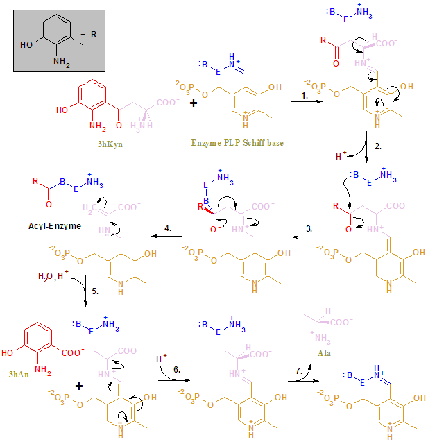
| The KYNU’s reaction mechanism. The color scheme is as follows: KYNU (blue), PLP (light brown), substrate names (green), inorganic molecules (dark brown), 3hAn’s moiety (red), Ala’s moiety (lavender) |
References
- PDB: 2HZP; Lima S, Khristoforov R, Momany C, Phillips RS (March 2007). “Crystal structure of Homo sapiens kynureninase”. Biochemistry. 46 (10): 2735–44. doi:10.1021/bi0616697. PMC 2531291. PMID 17300176.
- GRCh38: Ensembl release 89: ENSG00000115919 – Ensembl, May 2017
- GRCm38: Ensembl release 89: ENSMUSG00000026866 – Ensembl, May 2017
- “Human PubMed Reference:”. National Center for Biotechnology Information, U.S. National Library of Medicine.
- “Mouse PubMed Reference:”. National Center for Biotechnology Information, U.S. National Library of Medicine.
- Alberati-Giani D, Buchli R, Malherbe P, Broger C, Lang G, Köhler C, Lahm HW, Cesura AM (July 1996). “Isolation and expression of a cDNA clone encoding human kynureninase”. Eur. J. Biochem. 239 (2): 460–8. doi:10.1111/j.1432-1033.1996.0460u.x. PMID 8706755.
- Toma S, Nakamura M, Toné S, Okuno E, Kido R, Breton J, Avanzi N, Cozzi L, Speciale C, Mostardini M, Gatti S, Benatti L (May 1997). “Cloning and recombinant expression of rat and human kynureninase”. FEBS Lett. 408 (1): 5–10. doi:10.1016/S0014-5793(97)00374-8. PMID 9180257. S2CID 36265922.
- PDB: 3E9K; Lima S, Kumar S, Gawandi V, Momany C, Phillips RS (January 2009). “Crystal structure of the Homo sapiens kynureninase-3-hydroxyhippuric acid inhibitor complex: insights into the molecular basis of kynureninase substrate specificity”. J. Med. Chem. 52 (2): 389–96. doi:10.1021/jm8010806. PMID 19143568.
Further reading
- Lima S, Khristoforov R, Momany C, Phillips RS (2007). “Crystal structure of Homo sapiens kynureninase”. Biochemistry. 46 (10): 2735–2744. doi:10.1021/bi0616697. PMC 2531291. PMID 17300176.
- Heyes MP, Chen CY, Major EO, Saito K (1997). “Different kynurenine pathway enzymes limit quinolinic acid formation by various human cell types”. Biochem. J. 326 (2): 351–6. doi:10.1042/bj3260351. PMC 1218677. PMID 9291104.
- Rose JE, Behm FM, Drgon T, et al. (2010). “Personalized smoking cessation: interactions between nicotine dose, dependence and quit-success genotype score”. Mol. Med. 16 (7–8): 247–53. doi:10.2119/molmed.2009.00159. PMC 2896464. PMID 20379614.
- Zhang Y, Zhang KX, He X, et al. (2005). “[A polymorphism of kynureninase gene in a hypertensive candidate chromosomal region is associated with essential hypertension]”. Zhonghua Xin Xue Guan Bing Za Zhi. 33 (7): 588–91. PMID 16080802.
- Inada J, Okuno E, Kimura M, Kido R (1984). “Intracellular localization and characterization of 3-hydroxykynureninase in human liver”. Int. J. Biochem. 16 (6): 623–8. doi:10.1016/0020-711x(84)90031-4. PMID 6468727.
- Ubbink JB, Vermaak WJ, Bissbort SH (1991). “High-performance liquid chromatographic assay of human lymphocyte kynureninase activity levels”. J. Chromatogr. 566 (2): 369–75. doi:10.1016/0378-4347(91)80253-9. PMID 1939450.
- Maruyama K, Sugano S (1994). “Oligo-capping: a simple method to replace the cap structure of eukaryotic mRNAs with oligoribonucleotides”. Gene. 138 (1–2): 171–4. doi:10.1016/0378-1119(94)90802-8. PMID 8125298.
- Magni G, Amici A, Emanuelli M, et al. (2004). “Enzymology of NAD+ homeostasis in man”. Cell. Mol. Life Sci. 61 (1): 19–34. doi:10.1007/s00018-003-3161-1. PMID 14704851. S2CID 22041610.
- Strausberg RL, Feingold EA, Grouse LH, et al. (2002). “Generation and initial analysis of more than 15,000 full-length human and mouse cDNA sequences”. Proc. Natl. Acad. Sci. U.S.A. 99 (26): 16899–903. Bibcode:2002PNAS…9916899M. doi:10.1073/pnas.242603899. PMC 139241. PMID 12477932.
- Christensen M, Duno M, Lund AM, et al. (2007). “Xanthurenic aciduria due to a mutation in KYNU encoding kynureninase”. J. Inherit. Metab. Dis. 30 (2): 248–55. doi:10.1007/s10545-007-0396-2. PMID 17334708. S2CID 13295336.
- Walsh HA, Botting NP (2002). “Purification and biochemical characterization of some of the properties of recombinant human kynureninase”. Eur. J. Biochem. 269 (8): 2069–74. doi:10.1046/j.1432-1033.2002.02854.x. PMID 11985583.
- Suzuki Y, Yoshitomo-Nakagawa K, Maruyama K, et al. (1997). “Construction and characterization of a full length-enriched and a 5′-end-enriched cDNA library”. Gene. 200 (1–2): 149–56. doi:10.1016/S0378-1119(97)00411-3. PMID 9373149.
External links
- PDBe-KB provides an overview of all the structure information available in the PDB for Human Kynureninase
| Mitochondrial proteins |
|---|
| Metabolism: Protein metabolism, synthesis and catabolism enzymes |
|---|
| Hydrolases: carbon-carbon (EC 3.7) |
|---|
Leave a Reply