Peyer’s patches (or aggregated lymphoid nodules) are organized lymphoid follicles, named after the 17th-century Swiss anatomist Johann Conrad Peyer. They are an important part of gut associated lymphoid tissue usually found in humans in the lowest portion of the small intestine, mainly in the distal jejunum and the ileum, but also could be detected in the duodenum.
- Peyer, Johann Conrad (1677). Exercitatio Anatomico-Medica de Glandulis Intestinorum, Earumque Usu et Affectionibus [Anatomical-medical essay on the intestinal glands, and their function and diseases] (in Latin). Schaffhausen, Switzerland: Onophrius à Waldkirch.
- Reprinted as: Peyer, Johann Conrad (1681). Exercitatio Anatomico-Medica de Glandulis Intestinorum, Earumque Usu et Affectionibus (in Latin). Amsterdam, Netherlands: Henrik Wetstein.
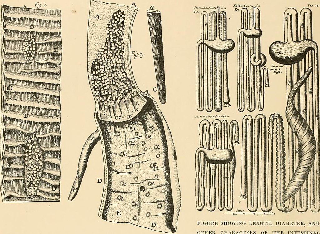
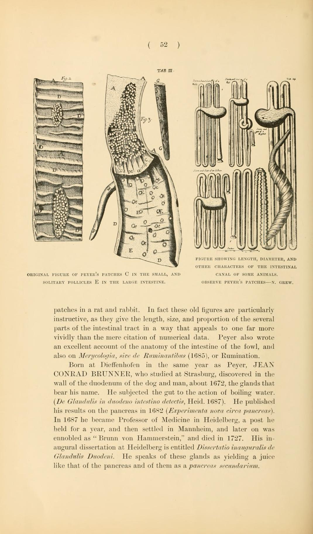
- Peyer referred to Peyer’s patches as plexus or agmina glandularum (clusters of glands). From (Peyer, 1681), p. 7: “Tenui a perfectiorum animalium Intestina accuratius perlustranti, crebra hinc inde, variis intervallis, corpusculorum glandulosorum Agmina sive Plexus se produnt, diversae Magnitudinis atque Figurae.” (I knew from careful study of more advanced animals, the intestines bear — often here and there, at various intervals — clusters of glandular small bodies or “plexuses” of diverse size and shape.) From p. 15: “(has Plexus seu agmina Glandularum voco)” (I call them “plexuses” or clusters of glands) He described their appearance. From p. 8: “Horum vero Plexuum facies modo in orbem concinnata; modo in Ovi aut Olivae oblongam, aliamve angulosam ac magis anomalam disposita figuram cernitur.” (But the configurations of these “plexuses” are arranged at one time in a circle; at another time, it is seen in an egg [shape] or an oblong olive [shape] or other faceted and more irregularly arranged shape.) Drawings of Peyer’s patches appear after pages 22 and 24.
- Zijlstra M, Auchincloss H, Loring JM, Chase CM, Russell PS, Jaenisch R (April 1992). “Skin graft rejection by beta 2-microglobulin-deficient mice”. The Journal of Experimental Medicine. 175 (4): 885–93. doi:10.1136/gut.6.3.225. PMC 1552287. PMID 18668776.
History
Peyer’s patches had been observed and described by several anatomists during the 17th century, but in 1677 Swiss anatomist Johann Conrad Peyer (1653–1712) described the patches so clearly that they were eventually named after him. However, Peyer regarded them as glands which discharged, into the small intestine, some substance which facilitated digestion. It was not until 1850 that the Swiss physician Rudolph Oskar Ziegler (1828–1881) suggested, after careful microscopic examination, that Peyer’s patches were actually lymph glands.
- ntestinorum, Earumque Usu et Affectionibus (in Latin). Amsterdam, Netherlands: Henrik Wetstein.
- Peyer referred to Peyer’s patches as plexus or agmina glandularum (clusters of glands). From (Peyer, 1681), p. 7: “Tenui a perfectiorum animalium Intestina accuratius perlustranti, crebra hinc inde, variis intervallis, corpusculorum glandulosorum Agmina sive Plexus se produnt, diversae Magnitudinis atque Figurae.” (I knew from careful study of more advanced animals, the intestines bear — often here and there, at various intervals — clusters of glandular small bodies or “plexuses” of diverse size and shape.) From p. 15: “(has Plexus seu agmina Glandularum voco)” (I call them “plexuses” or clusters of glands) He described their appearance. From p. 8: “Horum vero Plexuum facies modo in orbem concinnata; modo in Ovi aut Olivae oblongam, aliamve angulosam ac magis anomalam disposita figuram cernitur.” (But the configurations of these “plexuses” are arranged at one time in a circle; at another time, it is seen in an egg [shape] or an oblong olive [shape] or other faceted and more irregularly arranged shape.) Drawings of Peyer’s patches appear after pages 22 and 24.
- Haller, Albrecht von (1765). Elementa Physiologiae corporis humani [Elements of the physiology of the human body] (in Latin). Vol. 7. Bern, Switzerland: Societas Typographica. p. 35. Anatomists who mentioned Peyer’s patches included:
- Johann Theodor Schenck (1619–1671): Schenck, Johann Theodor (1672). Exercitationes Anatomicæ ad Usum Medicum Accommodatæ [Anatomical Exercises Suited to Medical Practice] (in Latin). Jena, (Germany): Johann Ludwig Neuenhahn. p. 334. Schenk thought that intestinal worms resided in Peyer’s patches and that the orifices of the patches were the worms’ mouths. From p. 334: “In canibus saepissime observavi non ad ventriculum … a praeter labente chylo sibi conveniens allicerent.” (In dogs, I very often noticed — not only near the stomach but also on the walls of their small intestines — flesh-colored or glandular blisters, [appearing] to swim one after another, [in] which, when we dissected [them], I observed some smooth reddish worms [vermium] living there in clusters [with] their heads facing towards the cavity of the intestines, in which part there were glands with orifices, [but] reversed, so that from there they obtained, from the chyle flowing past, nourishment [that was] suitable for them.)
- Jeremias Loss (1643–1684): Loss, Jeremias (1683). Dissertatio Medica de Glandulis in Genere [Medical Discourse on Glands in [Various] Species] (in Latin). Wittenberg, (Germany): Martin Schultz. p. 12. On page 12, Loss states that some glands are located “inter Membranas viscerum quorundam” (between the membranes of certain internal organs) “ … prout id in Glandulis Intestinorum satis manifestum est.” (as it is quite clear in the glands of the intestines), where “In Intestinis ita congregantur, interdum pauciores, interdum plures, ut areolas quasdam constituant: … ” (in the intestines there are thus gathered sometimes fewer [glands], sometimes more [glands], so that they form certain round patches.)
- Johannes Nicolaus Pechlin (1646–1706): Pechlin, Johannes Nicolaus (1672). De Purgantium Medicamentorum Facultatibus [On the Means of Medicinal Purges] (in Latin). Leiden and Amsterdam, Netherlands: Daniel, Abraham, and Adrian à Gaasbeek. p. 510. From p. 510: ” … ego tenuium glandularum glomeratum agmen esse ratus, … “ (… I considered the heaped cluster of fine glands, … )
- Martin Lister (ca. 1638–1712): Lister, Martin (23 June 1673). “A letter of Mr Lister dated May 21. 1673. in York, partly taking notice of the foregoing intimations, partly communicating some anatomical observations and experiments concerning the unalterable character of the whiteness of the chyle within the lacteous veins; together with divers particulars observed in the guts, especially several sorts of worms found in them”. Philosophical Transactions of the Royal Society of London. 8 (95): 6060–6065. Bibcode:1673RSPT….8.6060L. doi:10.1098/rstl.1673.0026. From p. 6062: “As 1. Glandulae miliares of the small Guts, which may also in some Animals be well call’d fragi-formes, from the figure of the one half of a Strawberry, and which yet I take to be Excretive glanduls, because Conglomerate.”
- Nehemiah Grew (1641–1712): Grew, Nehemiah (1681). The Comparative Anatomy of Stomachs and Guts Begun. Being Several Lectures Read before the Royal Society. In the Year, 1676. London, England: Self-published. p. 3. Grew called Peyer’s patches pancreas intestinale.
- There were many earlier names for Peyer’s patches:
- Todd, Robert Bentley, ed. (1859). The Cyclopædia of Anatomy and Physiology. Vol. 5. London, England: Longman, Brown, Green, Longmans, & Roberts. p. 356 footnote.
- Leidy, Joseph (1861). An Elementary Treatise on Human Anatomy. Philadelphia, Pennsylvania, USA: J.B. Lippincott & Co. p. 313 footnote.
- Ziegler, Rudolph Oskar (1850) Ueber die solitären und Peyerschen Follikel : Inaugural-Abhandlung, der medicinischen Facultät der Julius-Maximilians-Universität zu Würzburg vorgelegt [On solitary and Peyer’s follicles: Inaugural treatise, submitted to the medical faculty of the Julius-Maximilians-University of Würzburg] (in German) Würzburg, (Germany): Friederich Ernst Thein. From p. 37: “Ebensogross, wo nicht grösser ist die Aehnlichkeit der sogenannten Peyer’schen Drüsen und der Lymphdrüsen.” (Just as great, if not greater, is the resemblance between the so-called Peyer’s glands and the lymph glands.) From p. 38: ” … ja, man könnte selbst versucht sein, die letzteren für nichts als eine Art von zwischen den Wänden der Darmsschleimhaut eingebetteten Lymphdrüsen zu halten.” ( … indeed, one could even be tempted to regard the latter [i.e., the Peyer’s patches] as nothing but some type of lymph glands [which are] embedded between the walls of the intestinal mucosa.)
Structure
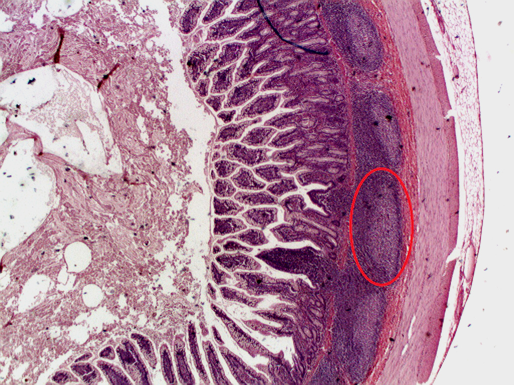
Peyer’s patches are observable as elongated thickenings of the intestinal mucosa measuring a few centimeters in length. About 100 are found in humans. Microscopically, Peyer’s patches appear as oval or round lymphoid follicles (similar to lymph nodes) located in the mucosa layer of the ileum and extend into the submucosa layer. The number of Peyer’s patches peaks at age 15–25 and then declines during adulthood. In the distal ileum, they are numerous and they form a lymphoid ring. At least 46% of Peyer’s patches are concentrated in the distal 25 cm of ileum in humans. It is important to note that there are large variations in size, shape, and distribution of Peyer’s patches from one individual to another one. In adults, B lymphocytes are seen to dominate the follicles’ germinal centers. T lymphocytes are found in the zones between follicles. Among the mononuclear cells, CD4+/CD25+ (10%) cells and CD8+/CD25+ (5%) cells are more abundant in Peyer’s patches than in the peripheral blood.
- Van Kruiningen HJ, West AB, Freda BJ, Holmes KA (May 2002). “Distribution of Peyer’s patches in the distal ileum”. Inflammatory Bowel Diseases. 8 (3): 180–5. doi:10.1097/00054725-200205000-00004. PMID 11979138. S2CID 22514793.
- Jung C, Hugot JP, Barreau F (September 2010). “Peyer’s Patches: The Immune Sensors of the Intestine”. International Journal of Inflammation. 2010: 823710. doi:10.4061/2010/823710. PMC 3004000. PMID 21188221.
- Zijlstra M, Auchincloss H, Loring JM, Chase CM, Russell PS, Jaenisch R (April 1992). “Skin graft rejection by beta 2-microglobulin-deficient mice”. The Journal of Experimental Medicine. 175 (4): 885–93. doi:10.1136/gut.6.3.225. PMC 1552287. PMID 18668776.
Peyer’s patches are characterized by the follicle-associated epithelium (FAE), which covers all lymphoid follicles. FAE differs from typical small intestinal villus epithelium: it has fewer goblet cells therefore mucus layer is thinner, and it is also characterized by the presence of specialized M cells or microfold cells, which provide uptake and transport of antigens from lumen. Moreover, basal lamina of follicle-associated epithelium is more porous compared to intestinal villus. Finally, follicle-associated epithelium is less permeable for ions and macromolecules, basically due to higher expression of tight junction proteins.
- Owen RL, Jones AL (February 1974). “Epithelial cell specialization within human Peyer’s patches: an ultrastructural study of intestinal lymphoid follicles”. Gastroenterology. 66 (2): 189–203. doi:10.1016/s0016-5085(74)80102-2. PMID 4810912.
- Onori P, Franchitto A, Sferra R, Vetuschi A, Gaudio E (May 2001). “Peyer’s patches epithelium in the rat: a morphological, immunohistochemical, and morphometrical study”. Digestive Diseases and Sciences. 46 (5): 1095–104. doi:10.1023/a:1010778532240. PMID 11341655. S2CID 34204173.
- Ermund A, Gustafsson JK, Hansson GC, Keita AV (2013). “Mucus properties and goblet cell quantification in mouse, rat and human ileal Peyer’s patches”. PLOS ONE. 8 (12): e83688. Bibcode:2013PLoSO…883688E. doi:10.1371/journal.pone.0083688. PMC 3865249. PMID 24358305.
- Takeuchi T, Gonda T (June 2004). “Distribution of the pores of epithelial basement membrane in the rat small intestine”. The Journal of Veterinary Medical Science. 66 (6): 695–700. doi:10.1292/jvms.66.695. PMID 15240945.
- Markov AG, Falchuk EL, Kruglova NM, Radloff J, Amasheh S (January 2016). “Claudin expression in follicle-associated epithelium of rat Peyer’s patches defines a major restriction of the paracellular pathway”. Acta Physiologica. 216 (1): 112–9. doi:10.1111/apha.12559. hdl:11701/6438. PMID 26228735. S2CID 13389571.
Function
Because the lumen of the gastrointestinal tract is exposed to the external environment, much of it is populated with potentially pathogenic microorganisms. Peyer’s patches thus establish their importance in the immune surveillance of the intestinal lumen and in facilitating production of the immune response within the mucosa.
Pathogenic microorganisms and other antigens entering the intestinal tract encounter macrophages, dendritic cells, B-lymphocytes, and T-lymphocytes found in Peyer’s patches and other sites of gut-associated lymphoid tissue (GALT). Peyer’s patches thus act for the gastrointestinal system much as the tonsils act for the respiratory system, trapping foreign particles, surveilling them, and destroying them. Peyer’s patches have adaptive immune capabilities through inducing selective apoptosis of B cells due CD122-targeted interleukin-2(IL-2) signaling. Additionally, the B cell population can be restored.
- Singh, Ayushi; Dhume, Kunal; Tejero, Joanne D.; Strutt, Tara M.; McKinstry, K. Kai (2020-07-29). “CD122-targetted IL-2 signals cause acute and selective apoptosis of B cells in Peyer’s Patches”. Scientific Reports. 10 (1): 12668. Bibcode:2020NatSR..1012668S. doi:10.1038/s41598-020-69632-5. ISSN 2045-2322. PMC 7391758. PMID 32728053.
Peyer’s patches are covered by a special follicle-associated epithelium that contains specialized cells called microfold cells (M cells) which sample antigen directly from the lumen and deliver it to antigen-presenting cells (located in a unique pocket-like structure on their basolateral side). Dendritic cells and macrophages can also directly sample the lumen by extending dendrites through transcellular M cell-specific pores. At the same time the paracellular pathway of follicle-associated epithelium is closed tightly to prevent penetration of antigens and continuous contact with immune cells. T cells, B-cells and memory cells are stimulated upon encountering antigen in Peyer’s patches. These cells then pass to the mesenteric lymph nodes where the immune response is amplified. Activated lymphocytes pass into the blood stream via the thoracic duct and travel to the gut where they carry out their final effector functions. The maturation of B-lymphocytes takes place in the Peyer’s patch.
- Lelouard H, Fallet M, de Bovis B, Méresse S, Gorvel JP (March 2012). “Peyer’s patch dendritic cells sample antigens by extending dendrites through M cell-specific transcellular pores”. Gastroenterology. 142 (3): 592–601.e3. doi:10.1053/j.gastro.2011.11.039. PMID 22155637.
- Bonnardel J, Da Silva C, Henri S, Tamoutounour S, Chasson L, Montañana-Sanchis F, Gorvel JP, Lelouard H (May 2015). “Innate and adaptive immune functions of peyer’s patch monocyte-derived cells”. Cell Reports. 11 (5): 770–84. doi:10.1016/j.celrep.2015.03.067. PMID 25921539.
- Diener M (January 2016). “Roadblock for antigens–take a detour via M cells”. Acta Physiologica. 216 (1): 13–4. doi:10.1111/apha.12595. PMID 26335934.
Clinical significance
Although important in the immune response, excessive growth of lymphoid tissue in Peyer’s patches is pathologic, as hypertrophy of Peyer’s patches has been closely associated with idiopathic intussusception.
Having too many or larger than normal Peyer’s patches is associated with an increased risk of prion diseases, and intussusception in children. A history of viral illness is a risk factor for enlarged or inflamed Peyer’s patches.
- MD, Steven M. Fiser (2022-08-30). The ABSITE Review (7th ed.). LWW. ISBN 978-1-9751-9029-3.
Intussusception may refer to:
- Intussusception (medical disorder)
- Intussusception (blood vessel growth)
- In a small study comparing the lungs of patients who had died from COVID-19 to those that had died from influenza A pneumonia (H1N1) to uninfected controls during autopsy; there was a significantly greater density of intussusceptive angiogenic features in the lungs of patients who had died from Covid-19 as compared to influenza A and the control group. The degree of intussusceptive angiogenic features in the lungs from the Covid-19 patients were also found to be greater as the length of hospitalization increased (which was not seen in the influenza or control groups). This suggests that increased or enhanced intussusceptive angiogenesis is seen in Covid-19 and may play a role in pathogenesis.
- Ackermann, Maximilian; Verleden, Stijn E.; Kuehnel, Mark; Haverich, Axel; Welte, Tobias; Laenger, Florian; Vanstapel, Arno; Werlein, Christopher; Stark, Helge; Tzankov, Alexandar; Li, William W.; Li, Vincent W.; Mentzer, Steven J.; Jonigk, Danny (21 May 2020). “Pulmonary Vascular Endothelialitis, Thrombosis, and Angiogenesis in Covid-19”. New England Journal of Medicine. 383 (2): 120–128. doi:10.1056/NEJMoa2015432. PMC 7412750. PMID 32437596.
- “Injury patterns in COVID-19 lungs”. Panta Rhei Study Group. Retrieved 6 July 2020.
- In a small study comparing the lungs of patients who had died from COVID-19 to those that had died from influenza A pneumonia (H1N1) to uninfected controls during autopsy; there was a significantly greater density of intussusceptive angiogenic features in the lungs of patients who had died from Covid-19 as compared to influenza A and the control group. The degree of intussusceptive angiogenic features in the lungs from the Covid-19 patients were also found to be greater as the length of hospitalization increased (which was not seen in the influenza or control groups). This suggests that increased or enhanced intussusceptive angiogenesis is seen in Covid-19 and may play a role in pathogenesis.
- Rectal prolapse#Internal rectal intussusception
The link goes to this page:
Intussusception is a medical condition in which a part of the intestine folds into the section immediately ahead of it. It typically involves the small bowel and less commonly the large bowel. Symptoms include abdominal pain which may come and go, vomiting, abdominal bloating, and bloody stool. It often results in a small bowel obstruction. Other complications may include peritonitis or bowel perforation. The cause in children is typically unknown; in adults a lead point is sometimes present. Risk factors in children include certain infections, diseases like cystic fibrosis, and intestinal polyps. Risk factors in adults include endometriosis, bowel adhesions, and intestinal tumors.
- Marsicovetere, P; Ivatury, SJ; White, B; Holubar, SD (February 2017). “Intestinal Intussusception: Etiology, Diagnosis, and Treatment”. Clinics in Colon and Rectal Surgery. 30 (1): 30–39. doi:10.1055/s-0036-1593429. PMC 5179276. PMID 28144210.
Salmonella typhi and poliovirus also target this section of the intestine.
- Pascall, C R; Stearn, E J; Mosley, J G (1980-07-05), “Short Reports”, British Medical Journal, vol. 281, no. 6232, p. 26, doi:10.1136/bmj.281.6232.26-a, PMC 1713722, PMID 7407483,
Unlike S hadar peritonitis, S typhi peritonitis is due to perforation of Peyer’s patches.
See also
References
Wikimedia Commons has media related to Peyer’s patches.
- Peyer, Johann Conrad (1677). Exercitatio Anatomico-Medica de Glandulis Intestinorum, Earumque Usu et Affectionibus [Anatomical-medical essay on the intestinal glands, and their function and diseases] (in Latin). Schaffhausen, Switzerland: Onophrius à Waldkirch.
- Reprinted as: Peyer, Johann Conrad (1681). Exercitatio Anatomico-Medica de Glandulis Intestinorum, Earumque Usu et Affectionibus (in Latin). Amsterdam, Netherlands: Henrik Wetstein.
- Peyer referred to Peyer’s patches as plexus or agmina glandularum (clusters of glands). From (Peyer, 1681), p. 7: “Tenui a perfectiorum animalium Intestina accuratius perlustranti, crebra hinc inde, variis intervallis, corpusculorum glandulosorum Agmina sive Plexus se produnt, diversae Magnitudinis atque Figurae.” (I knew from careful study of more advanced animals, the intestines bear — often here and there, at various intervals — clusters of glandular small bodies or “plexuses” of diverse size and shape.) From p. 15: “(has Plexus seu agmina Glandularum voco)” (I call them “plexuses” or clusters of glands) He described their appearance. From p. 8: “Horum vero Plexuum facies modo in orbem concinnata; modo in Ovi aut Olivae oblongam, aliamve angulosam ac magis anomalam disposita figuram cernitur.” (But the configurations of these “plexuses” are arranged at one time in a circle; at another time, it is seen in an egg [shape] or an oblong olive [shape] or other faceted and more irregularly arranged shape.) Drawings of Peyer’s patches appear after pages 22 and 24.
- Zijlstra M, Auchincloss H, Loring JM, Chase CM, Russell PS, Jaenisch R (April 1992). “Skin graft rejection by beta 2-microglobulin-deficient mice”. The Journal of Experimental Medicine. 175 (4): 885–93. doi:10.1136/gut.6.3.225. PMC 1552287. PMID 18668776.
- Haller, Albrecht von (1765). Elementa Physiologiae corporis humani [Elements of the physiology of the human body] (in Latin). Vol. 7. Bern, Switzerland: Societas Typographica. p. 35. Anatomists who mentioned Peyer’s patches included:
- Johann Theodor Schenck (1619–1671): Schenck, Johann Theodor (1672). Exercitationes Anatomicæ ad Usum Medicum Accommodatæ [Anatomical Exercises Suited to Medical Practice] (in Latin). Jena, (Germany): Johann Ludwig Neuenhahn. p. 334. Schenk thought that intestinal worms resided in Peyer’s patches and that the orifices of the patches were the worms’ mouths. From p. 334: “In canibus saepissime observavi non ad ventriculum … a praeter labente chylo sibi conveniens allicerent.” (In dogs, I very often noticed — not only near the stomach but also on the walls of their small intestines — flesh-colored or glandular blisters, [appearing] to swim one after another, [in] which, when we dissected [them], I observed some smooth reddish worms [vermium] living there in clusters [with] their heads facing towards the cavity of the intestines, in which part there were glands with orifices, [but] reversed, so that from there they obtained, from the chyle flowing past, nourishment [that was] suitable for them.)
- Jeremias Loss (1643–1684): Loss, Jeremias (1683). Dissertatio Medica de Glandulis in Genere [Medical Discourse on Glands in [Various] Species] (in Latin). Wittenberg, (Germany): Martin Schultz. p. 12. On page 12, Loss states that some glands are located “inter Membranas viscerum quorundam” (between the membranes of certain internal organs) “ … prout id in Glandulis Intestinorum satis manifestum est.” (as it is quite clear in the glands of the intestines), where “In Intestinis ita congregantur, interdum pauciores, interdum plures, ut areolas quasdam constituant: … ” (in the intestines there are thus gathered sometimes fewer [glands], sometimes more [glands], so that they form certain round patches.)
- Johannes Nicolaus Pechlin (1646–1706): Pechlin, Johannes Nicolaus (1672). De Purgantium Medicamentorum Facultatibus [On the Means of Medicinal Purges] (in Latin). Leiden and Amsterdam, Netherlands: Daniel, Abraham, and Adrian à Gaasbeek. p. 510. From p. 510: ” … ego tenuium glandularum glomeratum agmen esse ratus, … “ (… I considered the heaped cluster of fine glands, … )
- Martin Lister (ca. 1638–1712): Lister, Martin (23 June 1673). “A letter of Mr Lister dated May 21. 1673. in York, partly taking notice of the foregoing intimations, partly communicating some anatomical observations and experiments concerning the unalterable character of the whiteness of the chyle within the lacteous veins; together with divers particulars observed in the guts, especially several sorts of worms found in them”. Philosophical Transactions of the Royal Society of London. 8 (95): 6060–6065. Bibcode:1673RSPT….8.6060L. doi:10.1098/rstl.1673.0026. From p. 6062: “As 1. Glandulae miliares of the small Guts, which may also in some Animals be well call’d fragi-formes, from the figure of the one half of a Strawberry, and which yet I take to be Excretive glanduls, because Conglomerate.”
- Nehemiah Grew (1641–1712): Grew, Nehemiah (1681). The Comparative Anatomy of Stomachs and Guts Begun. Being Several Lectures Read before the Royal Society. In the Year, 1676. London, England: Self-published. p. 3. Grew called Peyer’s patches pancreas intestinale.
- ^ There were many earlier names for Peyer’s patches:
- Todd, Robert Bentley, ed. (1859). The Cyclopædia of Anatomy and Physiology. Vol. 5. London, England: Longman, Brown, Green, Longmans, & Roberts. p. 356 footnote.
- Leidy, Joseph (1861). An Elementary Treatise on Human Anatomy. Philadelphia, Pennsylvania, USA: J.B. Lippincott & Co. p. 313 footnote.
- ^ Ziegler, Rudolph Oskar (1850) Ueber die solitären und Peyerschen Follikel : Inaugural-Abhandlung, der medicinischen Facultät der Julius-Maximilians-Universität zu Würzburg vorgelegt [On solitary and Peyer’s follicles: Inaugural treatise, submitted to the medical faculty of the Julius-Maximilians-University of Würzburg] (in German) Würzburg, (Germany): Friederich Ernst Thein. From p. 37: “Ebensogross, wo nicht grösser ist die Aehnlichkeit der sogenannten Peyer’schen Drüsen und der Lymphdrüsen.” (Just as great, if not greater, is the resemblance between the so-called Peyer’s glands and the lymph glands.) From p. 38: ” … ja, man könnte selbst versucht sein, die letzteren für nichts als eine Art von zwischen den Wänden der Darmsschleimhaut eingebetteten Lymphdrüsen zu halten.” ( … indeed, one could even be tempted to regard the latter [i.e., the Peyer’s patches] as nothing but some type of lymph glands [which are] embedded between the walls of the intestinal mucosa.)
- Van Kruiningen HJ, West AB, Freda BJ, Holmes KA (May 2002). “Distribution of Peyer’s patches in the distal ileum”. Inflammatory Bowel Diseases. 8 (3): 180–5. doi:10.1097/00054725-200205000-00004. PMID 11979138. S2CID 22514793.
- Jung C, Hugot JP, Barreau F (September 2010). “Peyer’s Patches: The Immune Sensors of the Intestine”. International Journal of Inflammation. 2010: 823710. doi:10.4061/2010/823710. PMC 3004000. PMID 21188221.
- Owen RL, Jones AL (February 1974). “Epithelial cell specialization within human Peyer’s patches: an ultrastructural study of intestinal lymphoid follicles”. Gastroenterology. 66 (2): 189–203. doi:10.1016/s0016-5085(74)80102-2. PMID 4810912.
- Onori P, Franchitto A, Sferra R, Vetuschi A, Gaudio E (May 2001). “Peyer’s patches epithelium in the rat: a morphological, immunohistochemical, and morphometrical study”. Digestive Diseases and Sciences. 46 (5): 1095–104. doi:10.1023/a:1010778532240. PMID 11341655. S2CID 34204173.
- Ermund A, Gustafsson JK, Hansson GC, Keita AV (2013). “Mucus properties and goblet cell quantification in mouse, rat and human ileal Peyer’s patches”. PLOS ONE. 8 (12): e83688. Bibcode:2013PLoSO…883688E. doi:10.1371/journal.pone.0083688. PMC 3865249. PMID 24358305.
- Takeuchi T, Gonda T (June 2004). “Distribution of the pores of epithelial basement membrane in the rat small intestine”. The Journal of Veterinary Medical Science. 66 (6): 695–700. doi:10.1292/jvms.66.695. PMID 15240945.
- Markov AG, Falchuk EL, Kruglova NM, Radloff J, Amasheh S (January 2016). “Claudin expression in follicle-associated epithelium of rat Peyer’s patches defines a major restriction of the paracellular pathway”. Acta Physiologica. 216 (1): 112–9. doi:10.1111/apha.12559. hdl:11701/6438. PMID 26228735. S2CID 13389571.
- Singh, Ayushi; Dhume, Kunal; Tejero, Joanne D.; Strutt, Tara M.; McKinstry, K. Kai (2020-07-29). “CD122-targetted IL-2 signals cause acute and selective apoptosis of B cells in Peyer’s Patches”. Scientific Reports. 10 (1): 12668. Bibcode:2020NatSR..1012668S. doi:10.1038/s41598-020-69632-5. ISSN 2045-2322. PMC 7391758. PMID 32728053.
- Lelouard H, Fallet M, de Bovis B, Méresse S, Gorvel JP (March 2012). “Peyer’s patch dendritic cells sample antigens by extending dendrites through M cell-specific transcellular pores”. Gastroenterology. 142 (3): 592–601.e3. doi:10.1053/j.gastro.2011.11.039. PMID 22155637.
- Bonnardel J, Da Silva C, Henri S, Tamoutounour S, Chasson L, Montañana-Sanchis F, Gorvel JP, Lelouard H (May 2015). “Innate and adaptive immune functions of peyer’s patch monocyte-derived cells”. Cell Reports. 11 (5): 770–84. doi:10.1016/j.celrep.2015.03.067. PMID 25921539.
- Diener M (January 2016). “Roadblock for antigens–take a detour via M cells”. Acta Physiologica. 216 (1): 13–4. doi:10.1111/apha.12595. PMID 26335934.
- MD, Steven M. Fiser (2022-08-30). The ABSITE Review (7th ed.). LWW. ISBN 978-1-9751-9029-3.
- Pascall, C R; Stearn, E J; Mosley, J G (1980-07-05), “Short Reports”, British Medical Journal, vol. 281, no. 6232, p. 26, doi:10.1136/bmj.281.6232.26-a, PMC 1713722, PMID 7407483,
Unlike S hadar peritonitis, S typhi peritonitis is due to perforation of Peyer’s patches.
External links
Listen to this article (4 minutes)Duration: 4 minutes and 12 seconds.4:12
This audio file was created from a revision of this article dated 30 July 2019, and does not reflect subsequent edits.
(Audio help · More spoken articles)
| Organs of the lymphatic system |
|---|
| Anatomy of the gastrointestinal tract, excluding the mouth |
|---|
Leave a Reply