Category: Gastrointestinal System
-
Microfold cells (or M cells)
Microfold cells (or M cells) are found in the gut-associated lymphoid tissue (GALT) of the Peyer’s patches in the small intestine, and in the mucosa-associated lymphoid tissue (MALT) of other parts of the gastrointestinal tract. These cells are known to initiate mucosal immunity responses on the apical membrane of the M cells and allow for transport of microbes and particles across the epithelial cell layer from the gut lumen to the lamina…
-
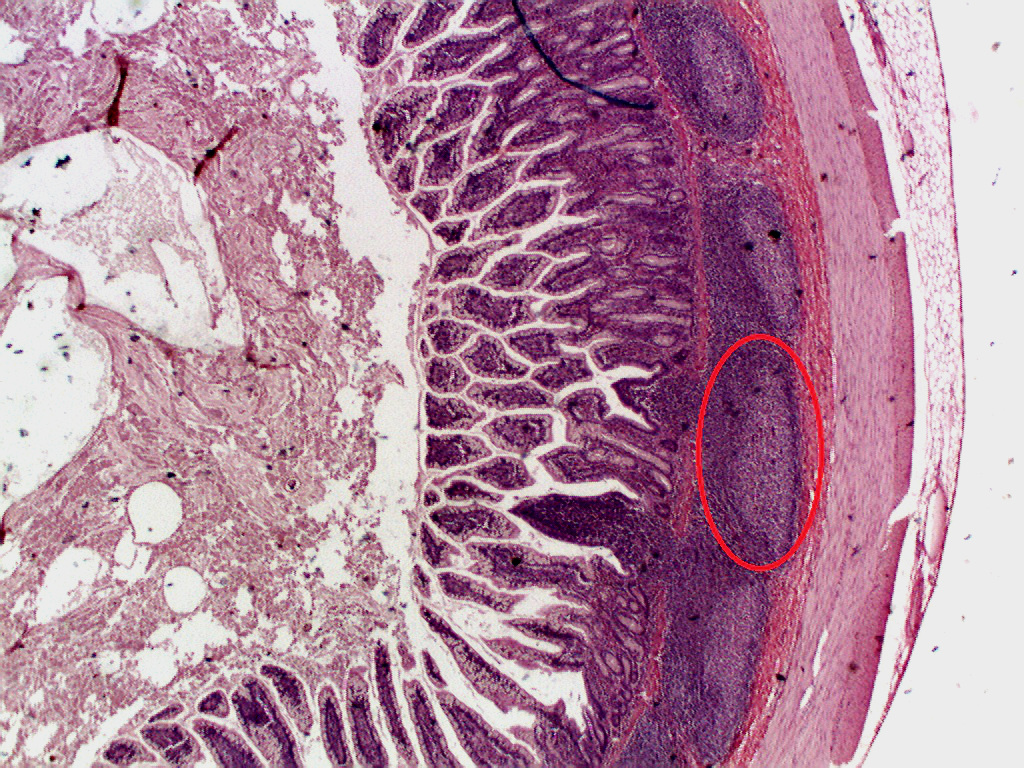
Peyer’s patches (aggregated lymphoid nodules)
Peyer’s patches (or aggregated lymphoid nodules) are organized lymphoid follicles, named after the 17th-century Swiss anatomist Johann Conrad Peyer. They are an important part of gut associated lymphoid tissue usually found in humans in the lowest portion of the small intestine, mainly in the distal jejunum and the ileum, but also could be detected in the duodenum. History Peyer’s patches had been observed and described by several anatomists…
-
The trefoil knot fold is a protein fold in which the protein backbone is twisted into a trefoil knot shape
“Shallow” knots in which the tail of the polypeptide chain only passes through a loop by a few residues are uncommon, but “deep” knots in which many residues are passed through the loop are extremely rare. Deep trefoil knots have been found in the SPOUT superfamily. including methyltransferase proteins involved in posttranscriptional RNA modification in all three domains of life, including bacterium Thermus thermophilus and…
-
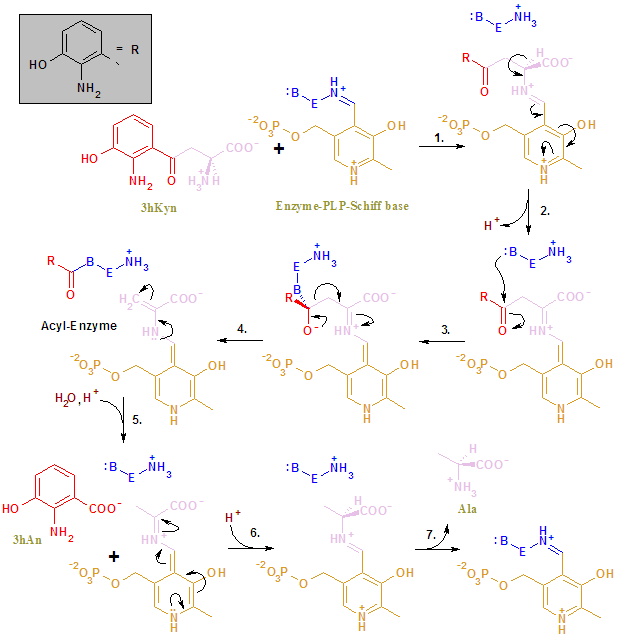
Kynureninase or L-Kynurenine hydrolase (KYNU) is part of the pathway for the catabolism of Trp and the biosynthesis of NAD cofactors from tryptophan (Trp).
Kynureninase or L-Kynurenine hydrolase (KYNU) (EC 3.7.1.3) is a PLP dependent enzyme that catalyses the cleavage of kynurenine (Kyn) into anthranilic acid (Ant). It can also act on 3-hydroxykynurenine (to produce 3-hydroxyanthranilate) and some other (3-arylcarbonyl)-alanines. Note: 3-Hydroxykynurenine is a metabolite of tryptophan, which filters UV light in the human lens. It is one of two pigments identified as responsible for the goldenrod crab spider‘s (Misumena vatia) yellow coloration. 3-Hydroxyanthranilic acid is an intermediate in the metabolism of tryptophan. It…
-

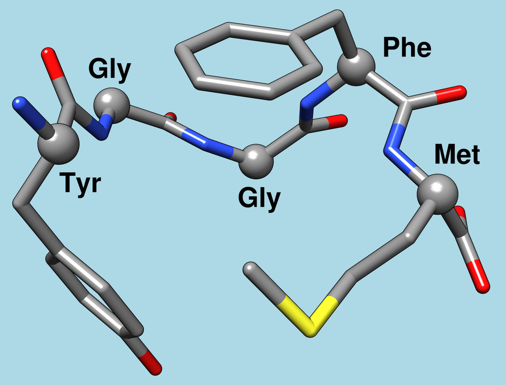
Leu-enkephalin and Met-enkephalin
Leu-enkephalin is an endogenous opioid peptide neurotransmitter with the amino acid sequence Tyr-Gly-Gly-Phe-Leu that is found naturally in the brains of many animals, including humans. It is one of the two forms of enkephalin; the other is met-enkephalin. The tyrosine residue at position 1 is thought to be analogous to the 3-hydroxyl group on morphine. Leu-enkephalin has agonistic actions at both the μ- and δ-opioid receptors, with significantly greater preference for the…
-
Enkephalins
An enkephalin is a pentapeptide involved in regulating nociception (pain sensation) in the body. The enkephalins are termed endogenous ligands, as they are internally derived and bind to the body’s opioid receptors. Discovered in 1975, two forms of enkephalin have been found, one containing leucine (“leu”), and the other containing methionine (“met”). Both are products of the proenkephalin gene. Endogenous opioid peptides There are three well-characterized families of opioid…
-
The pineal gland as an APUD organ
It is only in recent years that the pineal gland has emerged from being thought of as non-functional and unimportant. The rise from obscurity has been the result of the interest of investigators of multidisciplinary origins; such approaches, whilst clearly advancing understanding, also tend to leave knowledge fragmentary. In the last decade, a new neuroendocrine…
-
Pineal gland notes
The pineal gland, conarium, or epiphysis cerebri, is a small endocrine gland in the brain of most vertebrates. The pineal gland produces melatonin, a serotonin-derived hormone which modulates sleep patterns in both circadian and seasonal cycles. The shape of the gland resembles a pine cone, which gives it its name. The pineal gland is located in the epithalamus, near the center of the brain, between the two hemispheres, tucked in a groove where the two halves…
-
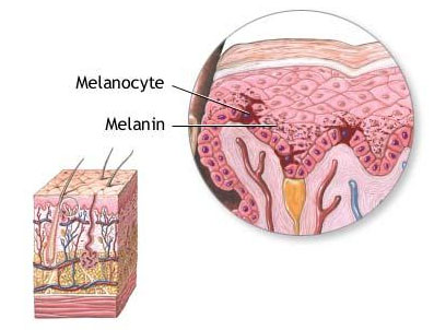
Cells in the APUD system may include melanocytes
Melanocytes are melanin-producing neural crest-derived cells located in the bottom layer (the stratum basale) of the skin’s epidermis, the middle layer of the eye (the uvea), the inner ear, vaginal epithelium, meninges, bones, and heart. Melanin is a dark pigment primarily responsible for skin color. Once synthesized, melanin is contained in special organelles called melanosomes which can be transported to nearby keratinocytes to induce pigmentation. Thus darker skin tones have more melanosomes present than lighter skin tones. Functionally, melanin serves as protection…
-
Cells in the APUD system may include Juxtaglomerular cells (JG cells), the renin producing cells in the kidney
Juxtaglomerular cells (JG cells), also known as juxtaglomerular granular cells are cells in the kidney that synthesize, store, and secrete the enzyme renin. They are specialized smooth muscle cells mainly in the walls of the afferent arterioles (and some in the efferent arterioles) that deliver blood to the glomerulus. In synthesizing renin, they play a critical role in the renin–angiotensin system and thus in autoregulation of the kidney. Juxtaglomerular cells secrete renin in…
-
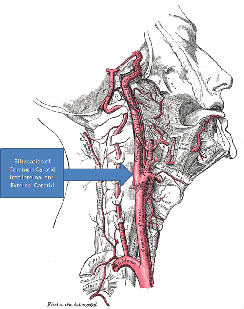
Carotid body glomus cells mediate essential reflex responses to arterial blood hypoxia
Glomus cells are the cell type mainly located in the carotid bodies and aortic bodies. Glomus type I cells are peripheral chemoreceptors which sense the oxygen, carbon dioxide and pH levels of the blood. When there is a decrease in the blood’s pH, a decrease in oxygen (pO2), or an increase in carbon dioxide (pCO2), the carotid bodies and the aortic bodies signal the dorsal…
-
The adrenal medulla is the principal site of the conversion of the amino acid tyrosine into the catecholamines; epinephrine, norepinephrine, and dopamine
The adrenal medulla (Latin: medulla glandulae suprarenalis) is part of the adrenal gland. It is located at the center of the gland, being surrounded by the adrenal cortex. It is the innermost part of the adrenal gland, consisting of chromaffin cells that secrete catecholamines, including epinephrine (adrenaline), norepinephrine (noradrenaline), and a small amount of dopamine, in response to stimulation by sympathetic preganglionic neurons. Structure The adrenal medulla consists of irregularly shaped cells…
-
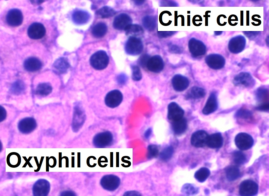
The chief cells of the parathyroid glands sense the amount of calcium in the blood, and release the calcium-increasing hormone parathyroid hormone (PTH) accordingly
Parathyroid chief cells (also called parathyroid principal cells or simply parathyroid cells) are one of the two cell types of the parathyroid glands, along with oxyphil cells. The chief cells are much more prevalent in the parathyroid gland than the oxyphil cells. It is perceived that oxyphil cells may be derived from chief cells at puberty, as they are not present at…
-
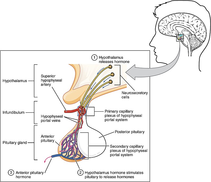
Adenohypophysis regulates several physiological processes, including stress, growth, reproduction, and lactation
A major organ of the endocrine system, the anterior pituitary (also called the adenohypophysis or pars anterior) is the glandular, anterior lobe that together with the posterior lobe (posterior pituitary, or the neurohypophysis) makes up the pituitary gland (hypophysis). The anterior pituitary regulates several physiological processes, including stress, growth, reproduction, and lactation. Proper functioning of the anterior pituitary and of the organs it regulates can often be ascertained via blood tests that measure hormone levels. Structure The pituitary gland sits in a…
-

Enterohepatic circulation
Enterohepatic circulation refers to the circulation of biliary acids, bilirubin, drugs or other substances from the liver to the bile, followed by entry into the small intestine, absorption by the enterocyte and transport back to the liver. Enterohepatic circulation is an especially important concept in the field of toxicology as many lipophilic xenobiotics undergo this process causing repeated liver damage. Enterohepatic circulation allows for recycling of metabolized and non-metabolized compounds,…