Protein structure
-
Structure of DNA repair protein XRCC4 aka X-ray repair cross-complementing protein 4
XRCC4 protein is a TETRAMER that resembles the shape of a DUMBBELL containing two globular ends separated by a long, thin stalk. The tetramer is composed of two dimers, and each dimer… Read more.
·
-
Pterion and Pteron Notes
The pterion is the region where the frontal, parietal, temporal, and sphenoid bones join. It is located on the side of the skull, just behind the temple. Structure The pterion is located in the temporal fossa, approximately 2.6 cm behind and 1.3 cm… Read more.
-
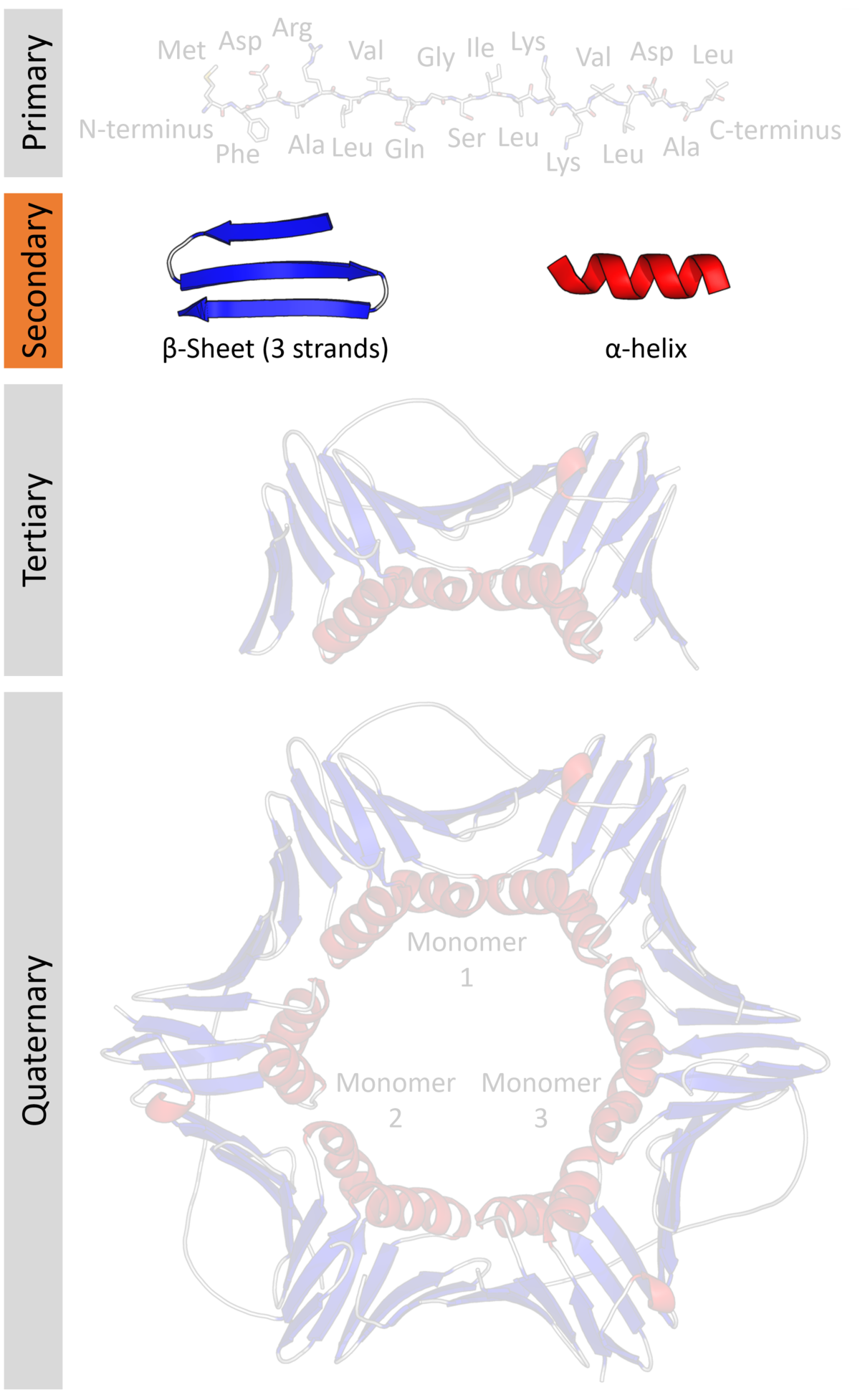
Protein secondary structure
Protein secondary structure is the three dimensional form of local segments of proteins. The two most common secondary structural elements are alpha helices and beta sheets, though beta turns and omega loops occur as well. Secondary structure elements typically spontaneously form as an intermediate before… Read more.
-
Turn (biochemistry)
For beta turns, see Beta turn. A turn is an element of secondary structure in proteins where the polypeptide chain reverses its overall direction. Definition According to one definition, see Rose et al. 1985 in… Read more.