Tag: Embryo
-

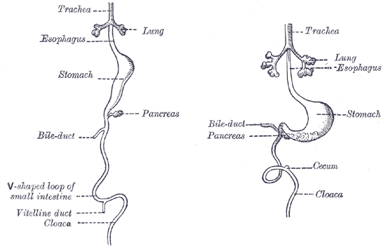
Vitelline duct connects the yolk sac to the small intestine. This duct obliterates when the embryo is about 6 weeks old. Complete failure of the duct to obliterate results in a fistula from the ileum to the umbilicus (vitelline fistula).
In the human embryo, the vitelline duct, also known as the vitellointestinal duct, the yolk stalk, the omphaloenteric duct, or the omphalomesenteric duct, is a long narrow tube that joins the yolk sac to the midgut lumen of the developing fetus. It appears at the end of the fourth week, when the yolk sac (also known as the umbilical vesicle) presents the appearance of a small pear-shaped vesicle. Function Obliteration Generally, the duct…
-

Albumen prints and egg whites…all the rage back in the day…and a few other things
The albumen print, also called albumen silver print, was published in January 1847 by Louis Désiré Blanquart-Evrard, and was the first commercially exploitable method of producing a photographic print on a paper base from a negative. It used the albumen found in egg whites to bind the photographic chemicals to the paper and became the dominant form of photographic positives from 1855 to the start…
-
Syncytin-2 also known as endogenous retrovirus group frd member 1
Syncytin-2 also known as endogenous retrovirus group FRD member 1 is a protein that in humans is encoded by the ERVFRD-1 gene.[5] This protein plays a key role in the implantation of human embryos in the womb.[6] This gene is conserved among all primates, with an estimated age of 45 million years. The receptor for this fusogenic env protein is MFSD2. The mouse…
-
Syncytiotrophoblast
Syncytiotrophoblast (from the Greek ‘syn’- “together”; ‘cytio’- “of cells”; ‘tropho’- “nutrition”; ‘blast’- “bud”) is the epithelial covering of the highly vascular embryonic placental villi, which invades the wall of the uterus to establish nutrient circulation between the embryo and the mother. It is a multi-nucleate, terminally differentiated syncytium, extending to 13 cm. It is the outer layer of the trophoblasts and actively invades the uterine wall, during implantation, rupturing maternal capillaries and thus establishing an interface between…
-
Cytotrophoblast
“Cytotrophoblast” is the name given to both the inner layer of the trophoblast (also called layer of Langhans) or the cells that live there. It is interior to the syncytiotrophoblast and external to the wall of the blastocyst in a developing embryo. The cytotrophoblast is considered to be the trophoblastic stem cell because the layer surrounding the blastocyst remains while daughter cells differentiate and proliferate…
-
What Is Metalloproteinase?
Metalloproteinase – the name alone screams “I’m here to ruin everything” – is a feral pack of enzymes armed with metal claws (zinc, mostly, because it’s the shiniest weapon in the elemental arsenal) that shred proteins like they’re auditioning for a slasher flick. These molecular psychopaths don’t just cut – they obliterate, turning the extracellular…
NOTES
- 🧬 Disease Table with Low Sodium Connection
- 🧂 Sodium Reduction and Sodium Replacement: A History of Reformulation and Exploding Diseases, Including Many Diseases Unheard of Before Deadly Sodium Policies
- 🧂 The DEADLY 1500 mg Sodium Recommendation predates the WHO’s formal global sodium reduction push by nearly a decade (and it’s even worse than that)
- 🧬 What Is Beta-Glucuronidase?
- When Sugar Was Salt: Crystalline Confusion and the Covenant of Sweetness
Tags
ADAM ASPARTAME Birds Blood Bones Brain Bugs Cancer Columba Cows crystallography Death Death cults Eggs Etymology Gastrin Gold Growth hormone History Hormones Insulin Liver Mere Perplexity Metal Monkey Business Mythology Paracetamol Plants Poison Pregnancy Protein Religion Reproduction Rocks Salt Slavery Snakes Sodium the birds and the bees Thiocyanate Tobacco Tylenol Underworld Venom zinc

