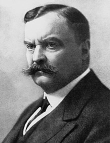
Erich Traub worked directly for Heinrich Himmler, head of the Schutzstaffel (SS), as the lab chief of the Nazis’ leading bio-weapons facility on Riems Island.
- Carroll, Michael (2004). Lab 257: The Disturbing Story of the Government’s Secret Germ Laboratory. New York: HarperCollins Publishers. ISBN 0-06-001141-6.
Note: Riems is home to the oldest virological research institution in the world, now called the Friedrich Loeffler Institute, which was built by Friedrich Loeffler in 1910. Loeffler, a professor at the University of Greifswald, ran filtration tests in 1898 and found that the cause of foot-and-mouth disease was not a bacterium, but a previously unknown class he called “the smallest of all organisms”. He had determined it to be a virus. After investigations showed that Loeffler had inadvertently infected the whole region of Greifswald with foot-and-mouth disease he moved to the safer location of his institute on the island of Riems in 1910. The Third Reich used the institute in Riems to research bioweapons. [Erhard Geißler: Hitler und die Biowaffen.. Lit, ISBN 3-8258-4077-8: S. 123 “… es sei gelungen, Rinder durch Versprühen eines in der Reichsforschungsanstalt Insel Riems hergestellten Viruspräparates … mit MKS zu infizieren.” (Google Books)] While East Germany controlled Riems approximately 800 people were working on vaccine research and development, today there are less than half that number. The population on the island is quite small. There are only 13 houses, five one- or two-family homes and eight apartment buildings, with a total of 62 residential units. Since 1997, the research complex is the headquarters of the Riemser Friedrich-Loeffler-Institute (FLI). The duties of the FLI include research on animal diseases, such as Bovine spongiform encephalopathy, foot-and-mouth disease and swine fever, and the development of preventive and protective measures against it, especially veterinary vaccines. As of 2006, Riems was working on a vaccine for the avian flu. By 2010, the Institute was to reduce their current locations Tübingen, Wusterhausen and Jena to just Jena and Riems. The total budget for the expansion work is some 150 million €. The construction needed to be handled carefully because of the historically rich old buildings. The former production plant for animal vaccines was successfully privatized as Riemser Arzneimittel AG. It has about 150 employees. After the 1990s the populated area in the western part of the island was freely accessible for some years. However, because of the renewed research work with viruses the island is again closed to the public. Quarantine stables and laboratories security levels are level 4. This means employees and visitors to the complex must change their clothes, and shower, when entering and exiting.
Traub was transported from the Soviet zone of Germany after World War II and taken to the United States in 1949 under the auspices of the United States government program Operation Paperclip, meant to exploit the post-war scientific knowledge in Germany, and deny it to the Soviet Union.
- Hunt, Hunt (1991). Secret Agenda: The United States Government, Nazi Scientists, and Project Paperclip, 1945 to 1990. New York: St.Martin’s Press. p. 340.
Career
Early career and war
During the 1930s, he studied on a fellowship at the Rockefeller Institute for Medical Research in Princeton, New Jersey mentored by Richard Shope, performing research on vaccines and viruses, including pseudorabies virus and lymphocytic choriomeningitis virus (LCM).
- Traub, Erich (22 March 1935). A filterable virus recovered from white mice. Science, volume 81. pp. 298–99.
- Traub E, Cultivation of Pseudorabies Virus, J Exp Med, 30 November 1933, 58(6), 663-81.
- Barthold SW, Introduction: microbes and the evolution of scientific fancy mice, ILAR J, 2008, 49(3), 265-71.
Note: Pseudorabies (also known as PRV, Aujeszky’s disease, infectious bulbar paralysis, or mad itch), is caused by a virus with icosahedral symmetry and belongs to the subfamily Alphaherpesvirinae within the family Herpesviridae. This subfamily has a wide host range and attacks the peripheral nervous system of the host. It was first described in 1813 in a situation where cattle and pigs shared a stable. In 1909 Weiss found that pigs are the reservoir host of the virus, and that, even though other species such as cattle, sheep, cats, dogs, goats, horses, raccoons, skunks, mice, and rats may transmit the disease, the virus completes its life cycle only in pigs.Aujeszky’s disease, usually called pseudorabies in the United States, is a viral disease in swine that has been endemic in most parts of the world. It is caused by Suid herpesvirus 1 (SuHV-1). Aujeszky’s disease is considered to be the most economically important viral disease of swine in areas where classical swine fever (hog cholera) has been eradicated.[Fenner, Frank J.; Gibbs, E. Paul J.; Murphy, Frederick A.; Rott, Rudolph; Studdert, Michael J.; White, David O. (1993). Veterinary Virology (2nd ed.). Academic Press, Inc. ISBN 978-0-12-253056-2.] Other mammals, such as cattle, sheep, goats, cats, dogs, and raccoons, are also susceptible. The disease is usually fatal in these animal species. Research on SuHV-1 in pigs has pioneered animal disease control with genetically modified vaccines. SuHV-1 is now used in model studies of basic processes during lytic herpesvirus infection, and for unravelling molecular mechanisms of herpesvirus neurotropism.[Mettenleiter (2008). “Molecular Biology of Animal Herpesviruses”. Animal Viruses: Molecular Biology. Caister Academic Press. ISBN 978-1-904455-22-6.][Sandri-Goldin, RM, ed. (2006). Alpha Herpesviruses: Molecular and Cellular Biology. Caister Academic Press. ISBN 978-1-904455-09-7.] In 1902, a Hungarian veterinarian, Aladár Aujeszky, demonstrated a new infectious agent in a dog, ox, and cat, and showed it caused the same disease in swine and rabbits. In the following decades the infection was found in several European countries, especially in cattle, where local intense pruritus (itching) is a characteristic symptom. And in the United States a well known disease in cattle called “mad itch” was concluded to be in fact Aujeszky’s disease.[Pomeranz L, Reynolds A, Hengartner C (2005). “Molecular Biology of Pseudorabies Virus: Impact on Neurovirology and Veterinary Medicine”. Microbiol Mol Biol Rev. 69 (3): 462–500. doi:10.1128/MMBR.69.3.462-500.2005. PMC 1197806. PMID 16148307.]
Note: Lymphocytic choriomeningitis (LCM) is a rodent-borne viral infectious disease that presents as aseptic meningitis, encephalitis or meningoencephalitis. Its causative agent is lymphocytic choriomeningitis mammarenavirus (LCMV), a member of the family Arenaviridae. The name was coined by Charles Armstrong in 1934.[Edward A. Beeman: Charles Armstrong, M.D.: A Biography, 2007 pp. 183ff. (also online here (PDF)] Lymphocytic choriomeningitis (LCM) is “a viral infection of the membranes surrounding the brain and spinal cord and of the cerebrospinal fluid”.[Lasker, Jill S. “Lymphocytic choriomeningitis”.] The name is based on the tendency of an individual to have abnormally high levels of lymphocytes during infection. Choriomeningitis is “cerebral meningitis in which there is marked cellular infiltration of the meninges, often with a lymphocytic infiltration of the choroid plexuses“.[ “choriomeningitis”] LCMV infection manifests itself in a wide range of clinical symptoms, and may even be asymptomatic for immunocompetent individuals.[CDC. “Information for Pet Owners: Reducing the Risk of Becoming Infected with LCMV from Pet Rodents”.] Onset typically occurs between one or two weeks after exposure to the virus and is followed by a biphasic febrile illness. During the initial or prodromal phase, which may last up to a week, common symptoms include fever, lack of appetite, headache, muscle aches, malaise, nausea, and/or vomiting. Less frequent symptoms include a sore throat and cough, as well as joint, chest, and parotid pain. The onset of the second phase occurs several days after recovery, and consists of symptoms of meningitis or encephalitis. Pathological findings during the first stage consist of leukopenia and thrombocytopenia. During the second phase, typical findings include elevated protein levels, increased leukocyte count, or a decrease in glucose levels of the cerebrospinal fluid).[United States. Div of Viral and Rickettsial Diseases, National Center for Infectious Diseases, CDC (August 2005). “Update: interim guidance for minimizing risk for human lymphocytic choriomeningitis virus infection associated with pet rodents”. MMWR Morb. Mortal. Wkly. Rep. 54 (32): 799–801. PMID 16107785.] Occasionally, a patient improves for a few days, then relapses with aseptic meningitis, or very rarely, meningoencephalitis. Patients with meningitis may have a stiff neck, fever, headache, myalgia, nausea and malaise. In some occasions, meningitis occurs without a prodromal syndrome. Meningoencephalitis is characterized by more profound neurological signs such as confusion, drowsiness, sensory abnormalities and motor signs. Under reported complications include myelitis, Guillain–Barré-type syndrome, cranial nerve palsies, transient or permanent hydrocephalus, sensorineural hearing loss, orchitis, arthritis and parotitis. LCMV infections have also been associated with pancreatitis, pneumonitis, myocarditis and pericarditis. The entire illness usually lasts 1 to 3 weeks,[The centre for Food Security and Public Health. Institute for International Co-operation and in Animal Biologics. Iowa State University. Ames, Iowa. 2010. http://www.cfsph.iastate.edu/Factsheets/pdfs/lymphocytic_choriomeningitis.pdf] nonetheless, temporary or permanent neurological damage is possible in all central nervous system infections, especially in cases of meningoencephalitis. Chronic infections have not been reported in humans and deaths rarely occur.[The centre for Food Security and Public Health. Institute for International Co-operation and in Animal Biologics. Iowa State University. Ames, Iowa. 2010. http://www.cfsph.iastate.edu/Factsheets/pdfs/lymphocytic_choriomeningitis.pdf]
During his stay in the United States, Traub and his wife were listed as members of the German American Bund, a pro-Nazi German-American club thirty miles west of Plum Island in Yaphank, Long Island, from 1934 to 1935.
- Carroll, Michael (2004). Lab 257: The Disturbing Story of the Government’s Secret Germ Laboratory. New York: HarperCollins Publishers. pp. 7–8. ISBN 0-06-001141-6.
Traub was a member of the Nazi NSKK, a motorist corps, from 1938 to 1942. The NSKK was declared a condemned, not a criminal organization at the Nuremberg trials.
- Carroll, Michael (2004). Lab 257: The Disturbing Story of the Government’s Secret Germ Laboratory. New York: HarperCollins Publishers. ISBN 0-06-001141-6.
Traub worked at the University of Giessen, Germany, from 1938 to 1942.
- Geissler, Erhard (1998). Conversion of BTW Warfare facilities: Lessons from German History. Springer; 1st edition. pp. 53–66. ISBN 978-0-7923-5250-1.
From 1942 to 1948, Traub worked as lab-chief at the Reich Research Institute for Virus Diseases of Animals (German: Reichsforschungsanstalt für Viruskrankheiten der Tiere) on Riems Island (German: Insel Riems), a German animal virus research institute in the Baltic sea, now named the Friedrich Loeffler Institute. The institute was headed by Prof. Dr. Otto Waldmann from 1919 to 1948, while Traub was vice-president.
- Geissler, Erhard (1998). Conversion of BTW Warfare facilities: Lessons from German History. Springer; 1st edition. pp. 53–66. ISBN 978-0-7923-5250-1.
The Institute at Riems Island was a dual use facility during the Second World War where at least some biological warfare experiments were conducted. It had been founded in 1909-10 to study foot-and-mouth disease in animals and by World War II employed about 20 scientists and a staff of 70-120. Hanns-Christoph Nagel, a veterinarian and biological warfare expert for the German Army, conducted experiments there, as did Traub.
- Geissler, Erhard (1998). Conversion of BTW Warfare facilities: Lessons from German History. Springer; 1st edition. pp. 53–66. ISBN 978-0-7923-5250-1.
Note: HFMD cases were first described clinically in Canada and New Zealand in 1957.[Wang, Jing (August 21, 2017). “Epidemiological characteristics of hand, foot, and mouth disease in Shandong, China, 2009–2016”. Scientific Reports. 7 (8900): 8900. Bibcode:2017NatSR…7.8900W. doi:10.1038/s41598-017-09196-z. PMC 5567189. PMID 28827733.] The disease was termed “Hand Foot and Mouth Disease”, by Thomas Henry Flewett, after a similar outbreak in 1960.[Alsop J, Flewett TH, Foster JR (December 1960). “”Hand-foot-and-mouth disease” in Birmingham in 1959″. British Medical Journal. 2 (5214): 1708–11. doi:10.1136/bmj.2.5214.1708. PMC 2098292. PMID 13682692.][Flewett TH, Warin RP, Clarke SK (January 1963). “‘Hand, foot, and mouth disease’ associated with Coxsackie A5 virus”. Journal of Clinical Pathology. 16 (1): 53–5. doi:10.1136/jcp.16.1.53. PMC 480485. PMID 13945538]
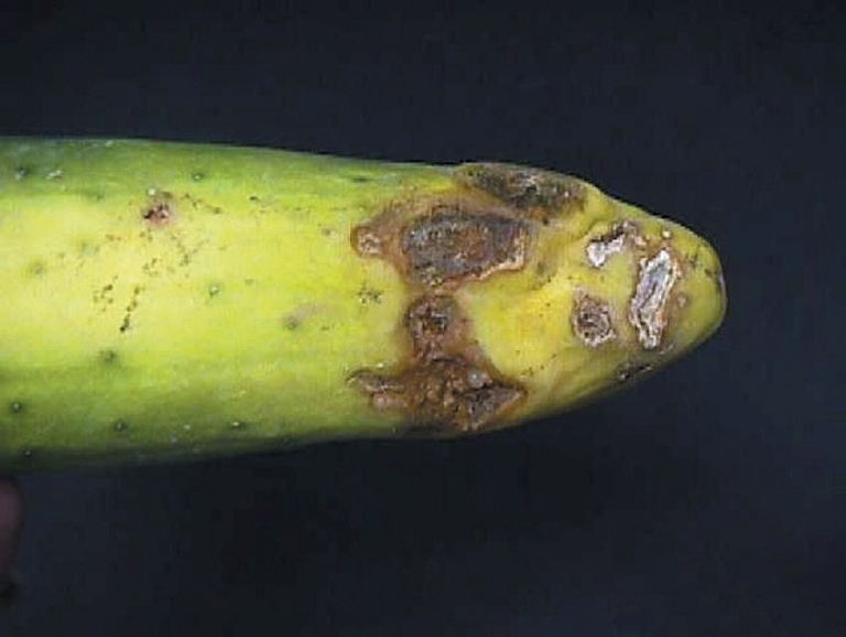
Note: Foot-and-mouth disease (FMD) or hoof-and-mouth disease (HMD) is an infectious and sometimes fatal viral disease that affects cloven-hoofed animals, including domestic and wild bovids.[Arzt, J.; Juleff, N.; Zhang, Z.; Rodriguez, L. L. (2011). “The Pathogenesis of Foot-and-Mouth Disease I: Viral Pathways in Cattle”. Transboundary and Emerging Diseases. 58 (4): 291–304. doi:10.1111/j.1865-1682.2011.01204.x. PMID 21366894][ Arzt, J.; Baxt, B.; Grubman, M. J.; Jackson, T.; Juleff, N.; Rhyan, J.; Rieder, E.; Waters, R.; Rodriguez, L. L. (2011). “The Pathogenesis of Foot-and-Mouth Disease II: Viral Pathways in Swine, Small Ruminants, and Wildlife; Myotropism, Chronic Syndromes, and Molecular Virus-Host Interactions”. Transboundary and Emerging Diseases. 58 (4): 305–326. doi:10.1111/j.1865-1682.2011.01236.x. PMID 21672184.] The virus causes a high fever lasting two to six days, followed by blisters inside the mouth and near the hoof that may rupture and cause lameness. Susceptible animals include cattle, water buffalo, sheep, goats, pigs,[Stenfeldt, C.; Pacheco, J.M.; Rodriguez, L.L.; Arzt, J. (2014). “Infection dynamics of foot-and-mouth disease virus in pigs using two novel simulated-natural inoculation methods”. Research in Veterinary Science. 96 (2): 396–405. doi:10.1016/j.rvsc.2014.01.009. PMID 24548596.][Stenfeldt, Carolina; Pacheco, Juan M.; Rodriguez, Luis L.; Arzt, Jonathan (2014). “Early Events in the Pathogenesis of Foot-and-Mouth Disease in Pigs; Identification of Oropharyngeal Tonsils as Sites of Primary and Sustained Viral Replication”. PLOS ONE. 9 (9): e106859. Bibcode:2014PLoSO…9j6859S. doi:10.1371/journal.pone.0106859. PMC 4153717. PMID 25184288.] antelope, deer, and bison. It has also been known to infect hedgehogs and elephants;[ “Canadian Food Inspection Agency – Animal Products – Foot-and-Mouth Disease Hazard Specific Plan”. Archived from the original on June 5, 2008.][McLauchlan, J. D.; Henderson, W. M. (1947). “The Occurrence of Foot-and-Mouth Disease in the Hedgehog under Natural Conditions”. The Journal of Hygiene. 45 (4): 474–479. doi:10.1017/s0022172400014194. PMC 2235060. PMID 18910334.] llamas and alpacas may develop mild symptoms, but are resistant to the disease and do not pass it on to others of the same species. In laboratory experiments, mice, rats, and chickens have been artificially infected, but they are not believed to contract the disease under natural conditions.[ “Canadian Food Inspection Agency – Animal Products – Foot-and-Mouth Disease Hazard Specific Plan”. Archived from the original on June 5, 2008.] Cattle, Asian and African buffalo, sheep, and goats can become carriers following an acute infection, meaning they are still infected with a small amount of virus but appear healthy. Animals can be carriers for up to 1–2 years and are considered very unlikely to infect other animals, although laboratory evidence suggests that transmission from carriers is possible.[Stenfeldt, Carolina; Arzt, Jonathan (28 February 2020). “The Carrier Conundrum; A Review of Recent Advances and Persistent Gaps Regarding the Carrier State of Foot-and-Mouth Disease Virus”. Pathogens. 9 (3): 167. doi:10.3390/pathogens9030167. PMC 7157498. PMID 32121072.][Arzt, Jonathan; Belsham, Graham J.; Lohse, Louise; Bøtner, Anette; Stenfeldt, Carolina (12 September 2018). “Transmission of Foot-and-Mouth Disease from Persistently Infected Carrier Cattle to Naive Cattle via Transfer of Oropharyngeal Fluid”. mSphere. 3 (5): e00365–18, /msphere/3/5/mSphere365–18.atom. doi:10.1128/mSphere.00365-18. PMC 6135961. PMID 30209130.] Humans are only extremely rarely infected by foot-and-mouth disease virus (FMDV). (Humans, particularly young children, can be affected by hand, foot, and mouth disease (HFMDV), which is often confused for FMDV. Similarly, HFMDV is a viral infection belonging to the Picornaviridae family, but it is distinct from FMDV. HFMDV also affects cattle, sheep, and swine.[“Hand, Foot, and Mouth Disease (HFMD)”. Centers for Disease Control and Prevention (CDC). 2019-02-22. Retrieved 28 August 2019.]) The virus responsible for FMD is an aphthovirus, foot-and-mouth disease virus. Infection occurs when the virus particle is taken into a cell of the host. The cell is then forced to manufacture thousands of copies of the virus, and eventually bursts, releasing the new particles in the blood. The virus is genetically highly variable,[Martinez-Salas E, Saiz M, Sobrino F (2008). “Foot-and-Mouth Disease Virus”. Animal Viruses: Molecular Biology. Caister Academic Press. pp. 1–38. ISBN 978-1-904455-22-6. Archived from the original on 2012-02-07. Retrieved 2007-08-10.] which limits the effectiveness of vaccination. The disease was first documented in 1870. The cause of FMD was first shown to be viral in 1897 by Friedrich Loeffler. He passed the blood of an infected animal through a Chamberland filter and found the collected fluid could still cause the disease in healthy animals. Humans can be infected with FMD through contact with infected animals, but this is extremely rare.[Capella, Giovanni Luigi (20 October 2001). “Foot and mouth disease in human beings”. The Lancet. 358 (9290): 1374. doi:10.1016/S0140-6736(01)06444-3. PMID 11684262. S2CID 26534737.] Some cases were caused by laboratory accidents. Because the virus that causes FMD is sensitive to stomach acid, it cannot spread to humans via consumption of infected meat, except in the mouth before the meat is swallowed. In the UK, the last confirmed human case occurred in 1966,[“Foot and Mouth Disease update: further temporary control zone established in Surrey”. Defra. 2007-08-14. Archived from the original on 2007-09-27. Retrieved 2007-08-14.][Jeffery, Simon (2001-11-23). “Foot and Mouth Disease”. The Guardian. London. Retrieved 2007-08-14.] and only a few other cases have been recorded in countries of continental Europe, Africa, and South America. Symptoms of FMD in humans include malaise, fever, vomiting, red ulcerative lesions (surface-eroding damaged spots) of the oral tissues, and sometimes vesicular lesions (small blisters) of the skin. According to a newspaper report, FMD killed two children in England in 1884, supposedly due to infected milk.[Jeffery, Simon (2001-11-23). “Foot and mouth ‘killed people in 1800s’”. The Guardian. London. Retrieved 2007-08-14.]
The institute was administered under the Innenministerium (Ministry of the Interior), which Reichsführer-SS Heinrich Himmler took over in 1943. The chain of command was Himmler, Dr. Leonardo Conti (Reich Health Leader), Kurt Blome, Waldmann, and then Traub. Traub specialized in viral and bacterial diseases. He was assisted by Anna Bürger, who was later also brought to the United States after the war, to work with the Navy’s biological warfare program.
- A Terrible Mistake: The Murder of Frank Olson and the CIA’s Secret Cold War Experiments – H.P.Albarelli – 1 July 2009 – ISBN 0-9777953-7-3
Note: Leonardo Conti (1900 – 1945) was the Reich Health Leader and an SS-Obergruppenführer in Nazi Germany. He was involved in the planning and execution of Action T4 that murdered hundreds of thousands adults and children with severe mental and physical handicaps. On 19 May 1945, after Germany’s surrender, Conti was imprisoned and in October hanged himself to avoid trial.Conti was born to a Swiss Italian father, Silvio, and a German mother, Nanna Pauli; his mother later became the Reich Midwifery Leader in Nazi Germany. He attended elementary school in Switzerland and the Friedrich Wilhelm Gymnasium in Berlin. In the summer of 1918, he volunteered for military service in the First World War with the Imperial German Army‘s 54th Field Artillery Regiment in Küstrin (today, Kostrzyn nad Odrą). However, he did not see any combat before the war ended in November.[Williams, Max (2015). SS Elite: The Senior Leaders of Hitler’s Praetorian Guard. Vol. 1. Fonthill Media LLC. ISBN 978-1-781-55433-3.] Conti was a staunch promoter of a public medical administration strongly controlled by the Nazi state. Under his leadership, local health offices were further expanded to allow for a genetic control and selection of the population in order to remove “weak” elements for the improvement of the German race, a doctrine known as eugenics. The various programmes were the basis for “racial hygiene” a lethal part of the Nazi philosophy. Conti worked with Dr. Karl Brandt to draft plans for the extermination of all Germany’s mental patients along with those suffering from severe physical handicaps. This program, known euphemistically as Action T4, is estimated to have killed over 200,000 adults and children between 1939 and 1945.[Childers, Thomas (2017). The Third Reich: A History of Nazi Germany. New York: Simon & Schuster. ISBN 978-1-45165-113-3.] Conti was initially placed in charge of this initiative but soon was replaced by Philip Bouhler.[Wistrich, Robert S. (4 July 2013). Who’s Who in Nazi Germany. Routledge. pp. 31–2. ISBN 978-1-136-41388-9.] Accordingly, he was co-responsible for the forced sterilization program, the racially motivated forced pregnancy interruptions, and ultimately the Action T4 euthanasia program. It is also undisputed that Conti participated in human experiments.[Leyh, Ernst-Alfred (2002) Leonardo Conti and the “ideologization of medicine during the Nazi dictatorship” (abstract). Universitätsbibliothek Heidelberg (in German).] Conti was also involved in the forensic investigation into the Katyn massacre, and received a detailed report, known as the Katyn Commission on the discovery from an international team of experts. On 19 May 1945, after Germany’s surrender, Conti was arrested by the British in Flensburg and was imprisoned and held as a witness for the Nuremberg trials. He would have been brought to the Doctors’ Trial for his involvement in Action T4. However, on 6 October 1945, Conti hanged himself in his Nuremberg cell.[Wistrich, Robert S. (4 July 2013). Who’s Who in Nazi Germany. Routledge. pp. 31–2. ISBN 978-1-136-41388-9.] On 1 May 1959, his estate was fined 3000 Deutsche Marks by the Berlin denazification tribunal.[Snyder, Louis (1994) [1976]. Encyclopedia of the Third Reich. Da Capo Press. ISBN 978-1-56924-917-8.]
- Conti, Leonardo . In: Wolfgang U. Eckart , Christoph Gradmann (eds.): Medical encyclopedia. From Antiquity to the Present . 3. Edition. Springer, Heidelberg 2006, ISBN 3-540-29584-4 , p. 84 f .
- Claude Cantini: Leonardo Conti. In: Historical Lexicon of Switzerland . February 21, 2007 .
- Eckhard Hansen, Florian Tennstedt (eds.) and others: Biographical dictionary on the history of German social policy from 1871 to 1945 . Volume 2: Social politicians in the Weimar Republic and under National Socialism 1919 to 1945. Kassel University Press, Kassel 2018, ISBN 978-3-7376-0474-1 , pp. 31–33 ( Online , PDF; 3.9 MB).
- Ernst-Alfred Leyh: “Health management”, “People’s destiny”, “Wehrmacht”. Leonardo Conti (1900-1945) and the ideologization of medicine under the Nazi dictatorship. (PDF) Diss. med. dent Man.], Medical Faculty Univ. Heidelberg 2002
- Joachim Lilla , Martin Döring, Andreas Schulz: extras in uniform. The members of the Reichstag 1933-1945. A biographical handbook. Including the Völkisch and National Socialist Reichstag deputies from May 1924. Droste, Düsseldorf 2004, ISBN 3-7700-5254-4 , pp. 81-83.
- Flavio Maggi: Un medico ticinese alla corte di Hitler. Leonardo Conti (1900-1945). Armando Dadò Editore , Locarno 199, ISBN 88-8281-031-3 .
- Thomas Maibaum: The leadership school of the German medical profession Alt-Rehse. University of Hamburg, dissertation in the Department of Medicine, 2007, p. 241 ff. Archive University of Hamburg (short biography.; PDF; 7.0 MB)
- Matthias Meusch: Conti Leonardo. In: Werner E. Gerabek , Bernhard D. Haage, Gundolf Keil , Wolfgang Wegner (ed.): Encyclopedia of medical history. De Gruyter, Berlin/ New York 2005, ISBN 3-11-015714-4 , p. 270.
- Literature by and about Leonardo Conti in the German National Library catalogue
- Newspaper article about Leonardo Conti in the historical press archives of the ZBW
- Documents on Leonardo Conti at the German Central Library for Economic Sciences
- Solveig Grothe: Puzzling Ending: Nazi Doctor in Wrong Grave ; Article at one day by Spiegel-Online.
- Ansgar Fabri, Jessica Thönnissen: Biography of Leonardo Conti In: Biographical Archive of Psychiatry (BIAPSY) , 2016.
- Leonardo Conti at ti.ch/can/oltreconfiniti
- Photos and illustrations:
- Estate of the Federal Archives N 2377
On orders from Himmler and Blome, the Deputy Reich Health Leader and head of the German biological warfare program, Traub worked on weaponizing foot-and-mouth disease virus, which has been reported to have been dispersed by aircraft onto cattle and reindeer in Russia.
- Glen Yeadon; John Hawkins (August 2008). The Nazi Hydra in America: Suppressed History of a Century. Progressive Press. p. 381. ISBN 978-0-930852-43-6.
Note: Kurt Blome (1894 – 1969) was a high-ranking Nazi scientist before and during World War II. He was the Deputy Reich Health Leader (Reichsgesundheitsführer) and Plenipotentiary for Cancer Research in the Reich Research Council. In his autobiography Arzt im Kampf (A Physician’s Struggle), he equated medical and military power in their battle for life and death. Blome was tried at the Doctors’ Trial in 1947 on charges of practicing euthanasia and conducting experiments on humans. He only admitted that he had been ordered in 1943 to experiment with plague vaccines on concentration camp prisoners. In reality, starting in 1943 he “assumed responsibility for all research into biological warfare sponsored by the Wehrmacht” and the SS.[Paul Maddrell, ‘Operation “Matchbox” and the Scientific Containment of the USSR’, in Peter Jackson & Jennifer Siegel (eds.)Intelligence and Statecraft: The Use and Limits of Intelligence in International Society. Praeger Publishers, 2005, p. 191.] Although he was acquitted of war crimes charges at the Nuremberg Doctors’ Trial, this was mainly due to the intervention of the United States as his earlier admissions were well known. It was generally accepted that he had indeed participated in chemical and biological warfare experiments on concentration camp inmates.[Alexander Cockburn and Jeffrey St. Clair, Whiteout: The CIA, Drugs and the Nazis. Verso, 1998, p. 148.] As Plenipotentiary for Cancer Research in the Third Reich, Blome had a longstanding interest in the “military use of carcinogenic substances” and cancer-causing viruses. According to Ute Deichmann‘s book Biologists under Hitler, in 1942 he became director of a unit affiliated with the Central Cancer Institute at the University of Posen (Poznań in Poland, annexed by Germany in 1939). Although he claimed that the work at this institute involved only ‘defensive’ measures against biological weapons, Heinrich Himmler, Hermann Goering, and Erich Schumann, head of the Wehrmacht’s Science Section, strongly supported the offensive use of chemical and biological weapons against Britain, the Soviet Union, and the United States. In 1943, Schumann wrote to Dr. Heinrich Kliewe, one of the Wehrmacht’s biological warfare experts that “in particular, America must be attacked simultaneously with various human and animal epidemic pathogens as well as plant pests.”[Ute Deichmann, Biologists under Hitler. Harvard University Press, 1996] According to Kliewe, plague, typhoid, cholera and anthrax were being developed as weapons, as well as a new “synthetic medium for the spread of these bacteria” which would allow them to remain virulent for eight to twelve weeks.[Ute Deichmann, Biologists under Hitler. Harvard University Press, 1996] Blome was arrested in Munich on 17 May 1945 by the United States Counter Intelligence Corps (an army intelligence service). He had no papers except his driving licence. After some weeks of custody, during which the CIC checked on his identity, Blome was taken to Kransberg Castle (a medieval castle north of Frankfurt). After his arrival at the castle a secret message was transmitted to Operation Alsos, an Anglo-American team of experts, tasked with investigating the state of German and Italian weapons technology towards the end of the war: In 1943 Blome was studying bacteriological warfare, although officially he was involved in cancer research, which was however only a camouflage. Blome additionally served as deputy health minister of the Reich. Would like you to send investigators?”[Alsos interrogation: in the Kurt Blome INSCOM dossier XE001248. Arrest reports: in Blome’s Nuremberg arrest file, RG 238, NARS.] It is believed that American intervention saved Blome from the gallows in exchange for information about biological warfare, nerve gas, and providing advice on to the American chemical and biological weapons programs.[Erhard Geissler, “Die Rolle deutscher Biowaffenexperten in der Zeit nach dem Zweiten Weltkrieg,” in Oehler-Klein & Roelcke,Vergangenheitspolitik in der universitaeren Medizin nach 1945 (Stuttgart: Franz Steiner, 2007), p. 101] In November 1947, two months after his Nuremberg acquittal, Blome was interviewed by four representatives from Camp Detrick, Maryland, including H.W. Batchelor, in which he “identified biological warfare experts and their location and described different methods of conducting biological warfare.”[Linda Hunt (April 1985). U.S. Coverup of Nazi Scientists. Bulletin of the Atomic Scientists. pp. 16–25.] Blome was hired by Sidney Gottlieb to work on the MK-Ultra program.[ Kinzer, Stephen (2019). Poisoner in Chief: Sidney Gottlieb and the CIA Search for Mind Control. New York: Henry Holt & Co.] In 1951, he was hired by the U.S. Army Chemical Corps under Project 63, one of the successors to Operation Paperclip, to work on chemical warfare. His file neglected to mention Nuremberg. Denied a visa by the U.S. Consul in Frankfurt, he was employed at European Command Intelligence Center at Oberursel, West Germany.[ George J. Annas; Michael A. Grodin (1 August 1995). The Nazi doctors and the Nuremberg Code: Human Rights in Human Experimentation. Oxford University Press. ISBN 978-0-19-510106-5.] He worked there on a never-declassified top secret project labeled in Blome’s foreign scientist case file as “Army, 1952, Project 1975”.[Annie Jacobsen: “Operation Paperclip: The Secret Intelligence Program that Brought Nazi Scientists to America” Little, Brown and Company, Inc. (2014) p. 364 ISBN 978-0316221047 (see also: “What Cold War CIA Interrogators Learned from the Nazis” The Daily Beast, 2. November 2014, (accessed Jan. 6, 2017) )] He was not arrested or charged with war crimes again after his acquittal at the Nuremberg Doctors’ Trial in 1947. He also continued to practice medicine in West Germany, and was active in politics as a member of the national-conservative German Party. He died in Dortmund in 1969.[Joachim Lilla: Extras in Uniform – The Members of the Reichstag 1933–1945 . Düsseldorf 2004, pp. 45–46.][better source needed] Works and related things:
- “Krebsforschung und Krebsbekämpfung”. Ziel und Weg. Die Gesundheitsführung Nr. 11 (1940) S. 406–412
- Arzt im Kampf: Erlebnisse und Gedanken. – Leipzig: Barth, 1942
- Literature by and about Kurt Blome in the German National Library catalogue
- About the behavior of bacteria in the electric current. Dissertation University of Rostock 1921.
- Since October 1939: Editor-in-chief of the monthly magazine Ziel und Weg. The health leadership.
- Cancer research and cancer control. In: Goal and way. The health leadership. No. 11, 1940, pp. 406-412.
- Doctor in battle: experiences and thoughts. Barth, Leipzig 1942 (listed in the Berlin Nazi bibliography since October 24, 1941).
- Institute for Contemporary History Munich-Berlin: Witnesses Online. ZS 830, Blome, Prof. Dr. Kurt (PDF; 12.3 MB). Minutes of Blome’s interrogations in 1946–1947.
- Jacobsen, Annie. Operation Paperclip: The Secret Intelligence Program that Brought Nazi Scientists to America. Little, Brown. 11 February 2014. ISBN 978-0-316-22105-4.
- McCoy, Alfred W. Science in Dachau’s Shadow: Hebb, Beecher, and the Development of CIA Psychological Torture and Modern Medical Ethics. Journal of the History of the Behavioral Sciences. Volume 143 (4), 2007.
- Entries of Kurt Blome in Rostock Matrikelportal
- Helge Dvorak: Biographical Encyclopedia of the German Fraternity. Volume I Politicians, Part 1: A–E. Heidelberg 1996, pp. 102–103.
- Ernst Kienast (ed.): The Greater German Reichstag 1938, IV. Election period. R. v. Decker’s Verlag, G. Schenck, June 1943 edition, Berlin.
- Joachim Lilla , Martin Döring, Andreas Schulz: extras in uniform. The members of the Reichstag 1933-1945. A biographical handbook. Including the Völkisch and National Socialist Reichstag deputies from May 1924. Droste, Düsseldorf 2004, ISBN 3-7700-5254-4 , pp. 45-46.
- Ernst Klee : The Person Encyclopedia of the Third Reich. Fischer paperback 2005, p. 54.
- Erhard Geißler : Biological weapons – not in Hitler’s arsenal. Biological and toxin warfare agents in Germany from 1915 to 1945. Munster 1998, ISBN 3-8258-2955-3 .
- Friedrich Hansen: Biological Warfare in the Third Reich. Campus-Verlag, Frankfurt am Main 1993 ( Review: Dirty Business . In: Der Spiegel . No. 43 , 1993, p. 227-233 ( online – October 25, 1993 ). )
- Thomas Maibaum: The leadership school of the German medical profession Alt-Rehse , University of Hamburg, Hamburg 2007, dissertation. PDF
- Gabriele Moser: From Deputy to “Reichsvollvollmachter” and Defendant at the Nuremberg Medical Trials: Dr. Kurt Blome and Cancer Research in National Socialist Germany , in: Wolfgang U. Eckart (ed.): Man, Medicine, and the State: The Human Body as an Object of Government Sponsored Medical Research in the 20th Century , Franz Steiner Verlag, Stuttgart 2006, pp. 199–220.
In 1944, Blome sent Traub to pick up a strain of Rinderpest virus in Turkey; upon his return, this strain proved inactive (nonvirulent) and therefore plans for a Rinderpest product were shelved.
- Carroll, Michael (2004). Lab 257: The Disturbing Story of the Government’s Secret Germ Laboratory. New York: HarperCollins Publishers. ISBN 0-06-001141-6.
Note: Rinderpest (also cattle plague or steppe murrain) was an infectious viral disease of cattle, domestic buffalo, and many other species of even-toed ungulates, including gaurs, buffaloes, large antelope, deer, giraffes, wildebeests, and warthogs.[Donald G. McNeil Jr. (27 June 2011). “Rinderpest, Scourge of Cattle, Is Vanquished”. The New York Times. Retrieved 28 June 2011.] The disease was characterized by fever, oral erosions, diarrhea, lymphoid necrosis, and high mortality. Death rates during outbreaks were usually extremely high, approaching 100% in immunologically naïve populations.[“Exotic animal diseases – Rinderpest”. .dpi.qld.gov.au. Archived from the original on March 30, 2010. Retrieved 2010-10-15.] Rinderpest was mainly transmitted by direct contact and by drinking contaminated water, although it could also be transmitted by air.[ “Rinderpest – the toll and treatment of a plague”. Food and Agriculture Organization (FAO). 1996. Archived from the original on 1997-06-09.] On 14 October 2010, the United Nations Food and Agriculture Organization (FAO) announced that field activities in the decades-long, worldwide campaign to eradicate the disease were ending, paving the way for a formal declaration in June 2011 of the global eradication of rinderpest.[ “UN ‘confident’ disease has been wiped out”. BBC News. 14 October 2010. Retrieved 14 October 2010.] On 25 May 2011, the World Organisation for Animal Health announced the free status of the last eight countries not yet recognized (a total of 198 countries were now free of the disease), officially declaring the eradication of the disease.[ “No More Deaths From Rinderpest” (Press release). World Organisation for Animal Health. Retrieved 25 May 2011.] In June 2011, the United Nations FAO confirmed the disease was eradicated, making rinderpest only the second disease in history to be fully wiped out (outside laboratory stocks), following smallpox.[McNeil Jr., Donald G. (27 June 2011). “Rinderpest, a Centuries-Old Animal Disease, Is Eradicated”. The New York Times.] In June 2019 the UK destroyed its stocks of rinderpest virus, held at the Pirbright Institute in Surrey, which were most of the world’s retained samples. This followed the completion of a digital record of the virus’s genetic code, thereby obviating the need to store samples as a protective resource in case the virus re-emerges. Researchers at Pirbright and the United Nations expressed a hope that the other samples in laboratories around the world will also be destroyed, totally eradicating the virus from the Earth.[ “Largest world stock of animal-killing virus destroyed by UK lab”. BBC News. 15 June 2019.] Rinderpest is believed to have originated in Asia, later spreading through the transport of cattle. The term Rinderpest is a German word meaning “cattle-plague”.[Donald G. McNeil Jr. (27 June 2011). “Rinderpest, Scourge of Cattle, Is Vanquished”. The New York Times. Retrieved 28 June 2011.][ Donald G. McNeil Jr. (15 October 2010). “Virus Deadly in Livestock Is No More, U.N. Declares”. The New York Times. Retrieved 15 October 2010.] The rinderpest virus (RPV) is closely related to the measles and canine distemper viruses.[Huygelen, C. (1997). “The immunization of cattle against rinderpest in eighteenth-century Europe”. Medical History. 41 (2): 182–196. doi:10.1017/s0025727300062372. PMC 1043905. PMID 9156464.] The measles virus possibly emerged from rinderpest as a zoonotic disease around 600 BC, a period that coincides with the rise of large human settlements.[Düx, Ariane; Lequime, Sebastian; Patrono, Livia Victoria; Vrancken, Bram; Boral, Sengül; Gogarten, Jan F.; Hilbig, Antonia; Horst, David; Merkel, Kevin; Prepoint, Baptiste; Santibanez, Sabine; Schlotterbeck, Jasmin; Suchard, Marc A.; Ulrich, Markus; Widulin, Navena; Mankertz, Annette; Leendertz, Fabian H.; Harper, Kyle; Schnalke, Thomas; Lemey, Philippe; Calvignac-Spencer, Sébastien (2020-06-19). “Measles virus and rinderpest virus divergence dated to the sixth century BCE”. Science. American Association for the Advancement of Science (AAAS). 368 (6497): 1367–1370. Bibcode:2020Sci…368.1367D. doi:10.1126/science.aba9411. ISSN 0036-8075. PMC 7713999. PMID 32554594. S2CID 219843735.][13] Rinderpest virus (RPV), a member of the genus Morbillivirus, is closely related to the measles and canine distemper viruses.[Huygelen, C. (1997). “The immunization of cattle against rinderpest in eighteenth-century Europe”. Medical History. 41 (2): 182–196. doi:10.1017/s0025727300062372. PMC 1043905. PMID 9156464.] Like other members of the Paramyxoviridae family, it produces enveloped virions, and is a negative-sense single-stranded RNA virus. The virus is particularly fragile and is quickly inactivated by heat, desiccation and sunlight.[ “Rinderpest”. Disease Facts. Institute for Animal Health. Archived from the original on June 26, 2009. Retrieved 2010-10-15.] Measles virus evolved from the then-widespread rinderpest virus most probably between the 11th and 12th centuries.[ Furuse, Yuki; Akira Suzuki; Hitoshi Oshitani (2010-03-04). “Origin of measles virus: divergence from rinderpest virus between the 11th and 12th centuries”. Virology Journal. 7: 52. doi:10.1186/1743-422X-7-52. ISSN 1743-422X. PMC 2838858. PMID 20202190.] The earliest likely origin is during the seventh century; some linguistic evidence exists for this earlier origin.[Griffin DE. In: Fields VIROLOGY. 5. Knipe DM, Howley PM, editor. Lippincott Williams & Wilkins; 2007. Measles Virus][McNeil W. Plagues and Peoples. New York: Anchor Press/Doubleday. 1976] In 2020 research on the measles virus has suggested a modified understanding of the evolution of rinderpest. Work on preserved older samples of measles (1912 and following) have been tested in various ways to determine the likely trajectory of the measles virus’ divergence from rinderpest. It is thought based on this study that the earliest date at which the divergence could have occurred is the sixth century BC.[Düx, Ariane; Lequime, Sebastian; Patrono, Livia Victoria; Vrancken, Bram; Boral, Sengül; Gogarten, Jan F.; Hilbig, Antonia; Horst, David; Merkel, Kevin; Prepoint, Baptiste; Santibanez, Sabine; Schlotterbeck, Jasmin; Suchard, Marc A.; Ulrich, Markus; Widulin, Navena; Mankertz, Annette; Leendertz, Fabian H.; Harper, Kyle; Schnalke, Thomas; Lemey, Philippe; Calvignac-Spencer, Sébastien (2020-06-19). “Measles virus and rinderpest virus divergence dated to the sixth century BCE”. Science. American Association for the Advancement of Science (AAAS). 368 (6497): 1367–1370. Bibcode:2020Sci…368.1367D. doi:10.1126/science.aba9411. ISSN 0036-8075. PMC 7713999. PMID 32554594. S2CID 219843735.] The disease is believed to have originated in Asia, later spreading through the transport of cattle. Other cattle epizootics are noted in ancient times: a cattle plague is thought to be one of the 10 plagues of Egypt described in the Hebrew Bible. By around 3,000 BC, a cattle plague had reached Egypt, and rinderpest later spread throughout the remainder of Africa, following European colonization.[Donald G. McNeil Jr. (15 October 2010). “Virus Deadly in Livestock Is No More, U.N. Declares”. The New York Times. Retrieved 15 October 2010.] In the 4th century, Roman writer Severus Sanctus Endelechius described rinderpest in his book, On the Deaths of Cattle.[Pastoret, Paul-Pierre; Yamanouchi, Kazuya; Mueller-Doblies*, Uwe; Rweyemamu, Mark M.; Horzinek, Marian; Barrett, Thomas (17 December 2005). “Rinderpest — an old and worldwide story: history to c. 1902”. Rinderpest and peste des petits ruminants : virus plagues of large and small ruminants. Academic Press. pp. 86–104. doi:10.1016/B978-012088385-1/50035-6. ISBN 978-0120883851.] Cattle plagues recurred throughout history, often accompanying wars and military campaigns. They hit Europe especially hard in the 18th century, with three long panzootics, which although varying in intensity and duration from region to region, took place in the periods of 1709–1720, 1742–1760, and 1768–1786.[Broad, J. (1983). “Cattle Plague in Eighteenth-Century England” (PDF). Agricultural History Review. 31 (2): 104–115. PMID 11620313. Retrieved 2013-09-17.]In the eighteenth century a deadly outbreak between 1769 and 1785 resulted in universal governmental action, but with somewhat divergent responses.[Van Roosbroeck, F (2019) Caring for Cows in a Time of Rinderpest: Non-academic Veterinary Practitioners in the County of Flanders, 1769–1785 The Journal of the Society for the Social History of Medicine 08/2019 Volume 32 Issue 3]The Dutch and the German principalities demanded quarantines and strict burial practices; England and large parts of Italy (the Papal States) saw slaughter of infected animals; in the Austrian Netherlands (Flanders) the response was inspection and precautionary slaughter coupled with compensation to the owners. There was no code of practice and no standard response. But for a hundred years thereafter in German-speaking countries there was intense focus on the problem of Rinderpest.[Sunseri, Thaddeus (2015). “The Entangled History of Sadoka (Rinderpest) and Veterinary Science in Tanzania and the Wider World, 1891–1901”. Bulletin of the History of Medicine. Project MUSE. 89 (1): 92–121. doi:10.1353/bhm.2015.0005. ISSN 1086-3176. PMID 25913464. S2CID 41707544.] In the early 18th century, the disease was seen as similar to smallpox, due to its analogous symptoms. The personal physician of the pope, Giovanni Maria Lancisi, recommended the destruction of all infected and exposed animals. This policy was not very popular and was used only sparingly in the first part of the century. Later, it was used successfully in several countries, although it was sometimes seen as too costly or drastic, and depended on a strong central authority to be effective (which was notably lacking in the Dutch Republic). Because of these downsides, numerous attempts were made to inoculate animals against the disease. These attempts met with varying success, but the procedure was not widely used and was no longer practiced at all in 19th-century Western or Central Europe. Rinderpest was an immense problem, but inoculation was not a valid solution. In many cases, it caused too many losses. Even more importantly, it perpetuated the circulation of the virus in the cattle population. The pioneers of inoculation did contribute significantly to knowledge about infectious diseases. Their experiments confirmed the concepts of those who saw infectious diseases as caused by specific agents, and were the first to recognize maternally derived immunity. Due to a very severe outbreak at the end of the 1760s, some of the best-known names in Dutch medicine became involved in the struggle against the disease. Several independent trials were begun, most notably by Pieter Camper in Groningen and Friesland. The results of his experiment in Friesland were encouraging, but they proved to be the exception; testing by others in the provinces of Utrecht and Friesland obtained disastrous results. As a result, the Frisian authorities concluded in 1769 that the cause of rinderpest was God’s displeasure with the sinful behavior of the Frisian people, and proclaimed 15 November a day of fasting and prayer. Interest in inoculation declined sharply across the country.[Huygelen, C. (1997). “The immunization of cattle against rinderpest in eighteenth-century Europe”. Medical History. 41 (2): 182–196. doi:10.1017/s0025727300062372. PMC 1043905. PMID 9156464.] In this climate of discouragement and scepticism, Geert Reinders, a farmer in the province of Groningen and a self-taught man, decided to continue the experiments. He collaborated with Wijnold Munniks, who had supervised earlier trials. They tried different inoculation procedures and a variety of treatments to lighten the symptoms, all of them without significant effect. Although they were not able to perfect the inoculation procedure, they did make some useful observations. Reinders resumed his experiments in 1774, concentrating on the inoculation of calves from cows that had recovered from rinderpest. He was probably the first to make practical use of maternally derived immunity. The detailed results of his trials were published in 1776 and reprinted in 1777. His inoculation procedure did not differ much from what had been used previously, except for the use of three separate inoculations at an early age. This produced far better results, and the publication of his work renewed interest in inoculation. For the period of 1777 to 1781, 89% of inoculated animals survived, compared to a 29% survival rate after natural infection. Apart from the Dutch Republic, the only other regions where inoculation was used to any significant level were northern Germany and Denmark. Experiments started in Mecklenburg during the epizootic of the late 1770s. “Insurance companies” were created which provided inoculation in special “institutes”. Although these were private initiatives, they were created with full encouragement from the authorities. Though neighboring states followed this practice with interest, the practice never caught on outside Mecklenburg; many were still opposed to inoculation. While some experimentation occurred in other countries (most extensively in Denmark), in the majority of European countries, the struggle against the disease was based on stamping it out. Sometimes this could be done with minimal sacrifices; at other times, it required slaughter at a massive scale.[Huygelen, C. (1997). “The immunization of cattle against rinderpest in eighteenth-century Europe”. Medical History. 41 (2): 182–196. doi:10.1017/s0025727300062372. PMC 1043905. PMID 9156464.] In 1917–18, William Hutchins Boynton (1881–1959), the chief veterinary pathologist with the Philippine Bureau of Agriculture, developed an early vaccine for rinderpest, based on treated animal organ extracts.[Boynton, W.H. (1917). “Preliminary report on the virulence of certain body organs in riderpest”. Philippine Agricultural Review. 10 (4): 410–433.][Boynton, W.H. (1918). “Use of organ extracts instead of virulent blood in immunization and hyperimmunization against rinderpest”. Philippine Journal of Science. 13 (3): 151–158.] In 1959, rinderpest vaccine was prepared at government laboratories in Abuko in The Gambia from the spleen of infected cattle. Walter Plowright worked on a vaccine for the RBOK strain of the rinderpest virus for about a decade, from 1956 to 1962.[Plowright, W.; Ferris, R. D. (1962). “Studies with rinderpest virus in tissue culture. The use of attenuated culture virus as a vaccine for cattle”. Res Vet Sci. 3: 172–182. doi:10.1016/S0034-5288(18)34916-6.] Plowright was awarded the World Food Prize in 1999 for developing a vaccine against a strain of rinderpest. In 1999, the FAO predicted that with vaccination, rinderpest would be eradicated by 2010.[EMPRES Transboundary Animal Diseases Bulletin No. 11 – Rinderpest”. Food and Agriculture Organization (FAO). 1923-07-20. Retrieved 2010-10-15.]
Post-war
Immediately after the war Traub was trapped in the Soviet zone of Allied occupied Germany. He was forced to work for the Soviets from his lab on Riems Island.
- Carroll, Michael (2004). Lab 257: The Disturbing Story of the Government’s Secret Germ Laboratory. New York: HarperCollins Publishers. pp. 10–11. ISBN 0-06-001141-6.
In July 1948, the British evacuated Erich Traub from Riems Island as a “high priority Intelligence target” since it was now in the Soviet Zone and they feared that Traub was assisting in their biological warfare program. Traub denied this, however, claiming that his only interest was foot-and-mouth disease in animals.
- Paul Maddrell, ‘Operation “Matchbox” and the Scientific Containment of the USSR’, in P. Jackson & J. Siegel (eds.), Intelligence and Statecraft: The Use and Limits of Intelligence in International Society ( Westport, CT : Praeger Publishers), (2005), pp. 173–206.
Traub was brought to the United States in 1949 under the auspices of the United States government program Operation Paperclip, meant to exploit scientific knowledge in Germany, and deny it to the Soviet Union.
- Hunt, Hunt (1991). Secret Agenda: The United States Government, Nazi Scientists, and Project Paperclip, 1945 to 1990. New York: St.Martin’s Press. p. 340.
From 1949 to 1953, he was associated with the Naval Medical Research Institute in Bethesda, Maryland.
- Geissler, Erhard (1998). Conversion of BTW Warfare facilities: Lessons from German History. Springer; 1st edition. pp. 53–66. ISBN 978-0-7923-5250-1.
Months into his Operation Paperclip contract, Traub was asked to meet with US scientists from Fort Detrick, the Army’s biological warfare headquarters, in Frederick, Maryland. As a noted German authority on viruses he was asked to consult on their animal disease program from a Biological Warfare perspective. Traub discussed work done at the Reich Research Institute for Virus Diseases of Animals on Riems Island during World War II for the Nazis, and work done after the war there for the Russians. Traub gave a detailed explanation of the secret operation at the Institute, and his activities there. This information provided the ground work for Fort Detrick’s offshore germ warfare animal disease lab on Plum Island.
- Carroll, Michael (2004). Lab 257: The Disturbing Story of the Government’s Secret Germ Laboratory. New York: HarperCollins Publishers. pp. 7–8. ISBN 0-06-001141-6.
Note: Fort Detrick is a United States Army Futures Command installation located in Frederick, Maryland. Fort Detrick was the center of the U.S. biological weapons program from 1943 to 1969. Since the discontinuation of that program, it has hosted most elements of the United States biological defense program.[https://mrdc.amedd.army.mil/assets/docs/media/USAMRDC-Handbook.pdf[bare URL PDF]] As of the early 2010s, Fort Detrick’s 1,200-acre (490 ha) campus supports a multi-governmental community that conducts biomedical research and development, medical materiel management, global medical communications and the study of foreign plant pathogens. The lab is known to research pathogens such as Ebola and smallpox.[“US army’s weapons lab shut down after inspection finds deadly viruses could escape”. The Independent. 2019-08-06. Retrieved 2020-06-21.] It is home to the U.S. Army Medical Research and Development Command (USAMRDC), with its bio-defense agency, the U.S. Army Medical Research Institute of Infectious Diseases (USAMRIID). It also hosts the Frederick National Laboratory for Cancer Research[“Frederick National Laboratory for Cancer Research”. Frederick National Laboratory for Cancer Research. National Cancer Institute, NIH, HHS. 2012-01-31. Retrieved 2023-03-29.] and is home to the National Interagency Confederation for Biological Research (NICBR),[ “National Interagency Confederation for Biological Research (NICBR)”. Detrick.army.mil. 2012-05-31. Retrieved 2012-07-26.] National Interagency Biodefense Campus (NIBC) and the National Center for Medical Intelligence (NCMI). In August 2019, its deadly germ research operations were shut down following serious safety violations, in particular relating to the disposal of dangerous materials.[Grady, Denise (2019-08-05). “Deadly Germ Research Is Shut Down at Army Lab Over Safety Concerns”. The New York Times. ISSN 0362-4331. Retrieved 2020-06-21.][DiGangi, Diana (2020-01-22). “Army germ lab shut down by CDC in 2019 had several ‘serious’ protocol violations that year”. WJLA. Retrieved 2020-06-21.] Fort Detrick is the largest employer in Frederick County, Maryland. Fort Detrick traces its roots to a small municipal airport established at Frederick, Maryland, in 1929. It was operated by a single person and the field was one of a string of emergency airfields between Cleveland, Ohio, and Washington, D.C., until 1938. The field was named in honor of squadron flight surgeon Major Frederick L. Detrick who served in France during World War I and died in June 1931 of a heart attack. During World War II, Camp Detrick and the USBWL became the site of intensive biological warfare (BW) research using various pathogens. This research was originally overseen by pharmaceuticals executive George W. Merck and for many years was conducted by Ira L. Baldwin, professor of bacteriology at the University of Wisconsin. Baldwin became the first scientific director of the labs. He chose Detrick Field for the site of this exhaustive research effort because of its balance between remoteness of location and proximity to Washington, D.C. – as well as to Edgewood Arsenal, the focal point of U.S. chemical warfare research. The Army’s Chemical Warfare Service was given responsibility and oversight for the effort that one officer described as “cloaked in the deepest wartime secrecy, matched only by … the Manhattan Project for developing the Atomic Bomb”.[Clendenin, Lt. Col. Richard M. (1968), Science and Technology at Fort Detrick, 1943–1968; Technical Information Division] After the defeat of Japan, the researchers working at Unit 731 were given immunity from prosecution. In return, director Shirō Ishii provided “8,000 slides of tissue from human and animal dissections” from the experiments, which were reportedly stored at Fort Detrick.[Dahl, Tracy (1983-05-26). “Japan’s Germ Warriors”. Washington Post. ISSN 0190-8286. Retrieved 2022-05-15.] From 1945 to 1955 under Project Paperclip and its successors, the U.S. government recruited over 1,600 German and Austrian scientists and engineers in a variety of fields such as aircraft design, missile technology and biological warfare. Among the specialists in the latter field who ended up working in the U.S. were Walter Schreiber, Erich Traub and Kurt Blome, who had been involved with medical experiments on concentration camp inmates to test biological warfare agents. Since Britain, France and the Soviet Union were also engaged in recruiting these scientists, the Joint Intelligence Objectives Agency (JIOA) wished to deny their services to other powers, and therefore altered or concealed the records of their Nazi past and involvement in war crimes.[Peter Knight, Conspiracy Theories in American History: An Encyclopedia, Volume One. ABC-CLIO, 2003.] On Veterans Day, November 11, 1969, President Richard M. Nixon asked the Senate to ratify the 1925 Geneva Protocol prohibiting the use of chemical and biological weapons. Nixon assured Fort Detrick its research would continue. On November 25, 1969, Nixon made a statement outlawing offensive biological research in the United States. Since that time any research done at Fort Detrick has been purely defensive in nature,[“History :: U.S. Army Fort Detrick”. home.army.mil. Retrieved 2021-12-15.] focusing on diagnostics, preventives and treatments for BW infections. This research is undertaken by the U.S. Army Medical Research Institute of Infectious Diseases (USAMRIID) which transitioned from the previous U.S. Army Medical Unit (USAMU) and was renamed in 1969. As he ended the offensive biological research done at Fort Detrick, Nixon pledged to make former laboratories and land available by the disestablishment of the offensive biological warfare program transferred to the U.S Department of Health and Human Services during the 1970s and later. The Frederick National Cancer Research and Development Center (now the Frederick National Laboratory for Cancer Research) was established in 1971 on a 69-acre (28 ha) parcel in Area A ceded by the installation.[“History :: U.S. Army Fort Detrick”. home.army.mil. Retrieved 2021-12-15.] In 1989 base researchers identified the Ebola virus in a monkey imported to the area from the Philippines.[Davis, Aaron, Michael E. Ruane and Nelson Hernandez, “Lab And Community Make For Uneasy Neighbors“, Washington Post, August 2, 2008, Pg. 10.] In the 1980s and 1990s, KGB disinformation agent Jakob Segal claimed that Fort Detrick was the site where the United States government “invented” HIV.[Johnson, I. (21 February 1992). “German scientist couple presses theory that AIDS was created at Fort Detrick”. Baltimore Sun. Retrieved 25 January 2017.]USAMRIID had been the principal consultant to the FBI on scientific aspects of the 2001 Anthrax Attacks, which had infected 22 people and killed five.[“Anthrax scientist commits suicide as FBI closes in”. Associated Press. Archived from the original on August 5, 2008. Retrieved 2008-08-01.] While assisting with the science from the beginning, it also soon became the focus of the FBI’s investigation of possible perpetrators (see Steven Hatfill). In July 2008, a top U.S. biodefense researcher at USAMRIID committed suicide just as the FBI was about to lay charges relating to the incidents. The scientist, Bruce Edwards Ivins, who had worked for 18 years at USAMRIID, had been told about the impending prosecution. The FBI’s identification of Ivins in August 2008 as the Anthrax Attack perpetrator remains controversial and several independent government investigations which will address his culpability are ongoing. Although the anthrax preparations used in the attacks were of different grades, all of the material derived from the same bacterial strain. Known as the Ames strain, it was first researched at USAMRIID. The Ames strain was subsequently distributed to at least fifteen bio-research labs within the U.S. and six locations overseas. In June 2008 the Environmental Protection Agency said it planned to add the base to the Superfund list of the most polluted places in the country.[Davis, Aaron, Michael E. Ruane and Nelson Hernandez, “Lab And Community Make For Uneasy Neighbors“, Washington Post, August 2, 2008, Pg. 10.] On 9 April 2009, “Fort Detrick Area B Ground Water” was added to the list which currently includes 18 other sites within Maryland. The Forest Glen Annex of the Walter Reed Army Medical Center in Silver Spring, Maryland was transferred to the command of Fort Detrick in 2008 as a result of the Base Realignment and Closure process.[“Fort Detrick 2010 Post Guide” (PDF). 2010. p. 6. Archived from the original (PDF) on 10 October 2011. Retrieved 26 July 2014.] In 2020, a conspiracy theory regarding COVID-19 arose that alleged that the SARS-CoV-2 virus was developed by the United States Army at Fort Detrick.[Helen Davidson (20 January 2021). “China revives conspiracy theory of US army link to Covid”. The Guardian.] This allegation has been promoted by Chinese government officials, most notably Ministry of Foreign Affairs spokesman Zhao Lijian, who has called for an inspection of the facility, although the allegation remains baseless.[Ang, Katerina; Taylor, Adam (May 28, 2021). “As U.S. calls for focus on covid origins, China repeats speculation about U.S. military base”. The Washington Post. Retrieved 29 May 2021.] A petition organized by the Chinese Communist Party-owned tabloid Global Times urging the WHO to investigate Fort Detrick for COVID origins reportedly amassed 25 million signatures.[Levin, Dan (25 August 2021). “Florida Sees Worst of Pandemic So Far”. The New York Times. Retrieved 29 August 2021.] During an inspection by the Centers for Disease Control and Prevention (CDC) of the United States Army Medical Research Institute of Infectious Diseases (USAMRIID) BSL-3 and BSL-4 laboratories at Ft Detrick in June 2019 six violations including two breaches of containment were identified. The inspection was followed up by a letter of concern from the CDC on July 12, 2019 and then a cease and desist order on July 15, 2019.[“CDC Inspection Findings Reveal More about Fort Detrick Research Suspension”. web.archive.org. 27 November 2019. Archived from the original on 2019-11-27. Retrieved 28 August 2021.] Following the cease and desist order from the CDC the USAMRIID laboratories at the base were shut down in August 2019. The announcement to resume operations on a “limited scale” was made on November 25, 2019. The CDC cited “national security reasons” as the reason for not informing the public about its decision.[ Grady, Denise (2019-08-05). “Deadly Germ Research Is Shut Down at Army Lab Over Safety Concerns”. The New York Times. ISSN 0362-4331. Retrieved 2020-06-21.] The two breaches reported to the CDC by USAMRIID staff demonstrated failures of biosafety level 3 and 4 protocols in the Army laboratory to “implement and maintain containment procedures sufficient to contain select agents or toxins”.[ “CDC Inspection Findings Reveal More about Fort Detrick Research Suspension”. web.archive.org. 27 November 2019. Archived from the original on 2019-11-27. Retrieved 28 August 2021.] After approximately eight months of closure and restrictions, the USAMRIID BSL-4 lab had been authorized to resume full operational status by April 2020, to the applause of Maryland lawmakers including Senator Ben Cardin, who stated “it is a relief to have USAMRIID fully operational with the current COVID-19 outbreak”‘.[Mongilio, Heather. “Fort Detrick laboratory restored to full operations after being shut down by CDC”. The Frederick News-Post. Retrieved 28 August 2021.][“Maryland lawmakers applaud CDC’s decision to reopen Fort Detrick facility”. Homeland Preparedness News. 1 April 2020. Retrieved 28 August 2021.] Each branch of the U.S. military is represented among Fort Detrick’s 7,800 military, federal and contractor employees. Four cabinet-level agencies are represented by activities on the garrison: The U.S. Department of Homeland Security, the U.S. Department of Agriculture, the U.S. Department of Health and Human Services, and the U.S. Department of Defense. The offices and laboratories include the Agriculture Department’s Foreign Disease and Weed Science Research Institute, the National Cancer Institute, the Naval Medical Logistics Command and the Telemedicine and Advanced Technology Research Center. Currently under construction is a biotechnology campus that will house civilian and military research centers including units of the Centers for Disease Control and Prevention (CDC), the National Institute of Allergy and Infectious Diseases (NIAID), as well as USAMRIID.[Wood, David, “Variety Of Research Carried Out At Fort Detrick“, Baltimore Sun, August 2, 2008.]
Traub publicly published research from his time in the United States reports disease research not directly related to weaponization. In 1951, he published a report for the Naval Medical Research Institute on Newcastle Disease virus in chicken and mammalian blood cells. Two years later, he published a paper for the Navy on the mechanisms of immunity in chickens to Newcastle and the possible role of cellular factors.
- Erich Traub, “Studies on the In-Vitro Multiplication of Newcastle Disease Virus in Chicken Blood.” Naval Medical Research Institute, National Naval Medical Center, 1951.
- Erich Traub, “Studies in the Mechanism of Immunity of Chickens to Newcastle Disease Virus.” Naval Medical Research Institute, National Naval Medical Center, 1953.
Also in 1953, he published another paper for the Navy with Worth I. Capps on the foot-and-mouth disease virus and methods for rapid adaptation.
- Erich Traub and Worth I. Capps, “Experiments with Chicken Embryo-Adapted Foot-and-Mouth Disease Virus and a Method for the Rapid Adaptation.” Naval Medical Research Institute, National Naval Medical Center, 1953.
Traub served as an expert on foot-and-mouth disease for the FAO of the UN in Bogota, Colombia, from 1951 to 1952, in Tehran, Iran, from 1963 to 1967, and in Ankara, Turkey, from 1969 to 1971.
Return to Germany

After working on biological research for the U.S. Navy from 1949 to 1953, Traub returned to Germany and founded a new branch of the Loeffler Institut in Tübingen, and headed it from 1953 to 1963.
- Friedrich-Loeffler-Institute, Federal Research Institute for Animal Health Archived 5 February 2012 at the Wayback Machine, History: Isle of Riems
In 1960, Traub resigned as Tübingen’s director due to the scandal related to accusations of financial embezzlement. He continued with limited lab research for three more years, but then ended his career at Tübingen.
- Carroll, Michael (2004). Lab 257: The Disturbing Story of the Government’s Secret Germ Laboratory. New York: HarperCollins Publishers. pp. 10–11. ISBN 0-06-001141-6.
In 1964, Traub published a study for the Army Biological labs in Frederick, Maryland on Eastern Equine Encephalomyeltitis (EEE) immunity in white mice and its relationship to Lymphocytic choriomeningitis (LCM), which had long been a research interest of his.
- Eric Traub, “Immunity of White Mice to EEE-VIrus.” Report No. 8, Army Biological Labs, Frederick, MD, 1964.
Note: Eastern equine encephalitis (EEE), commonly called Triple E or sleeping sickness (not to be confused with African trypanosomiasis), is a disease caused by a zoonotic mosquito vectored Togavirus that is present in North, Central, and South America, and the Caribbean. EEE was first recognized in Massachusetts, United States, in 1831, when 75 horses died mysteriously of viral encephalitis. Epizootics in horses have continued to occur regularly in the United States. It can also be identified in donkeys and zebras. Due to the rarity of the disease, its occurrence can cause economic impact beyond the cost of horses and poultry.[ “Eastern Equine Encephalomyelitis” (PDF). United States Department of Agriculture Animal and Plant Health Inspection Service. United States Department of Agriculture (USDA). Archived from the original (PDF) on 3 February 2017. Retrieved 1 May 2017.] EEE is found today in the eastern part of the United States and is often associated with coastal plains. It can most commonly be found in East Coast and Gulf Coast states.[“Eastern Equine Encephalitis”. Centers for Disease Control and Prevention (CDC). Retrieved 30 April 2017.] In Florida, about one to two human cases are reported a year, although over 60 cases of equine encephalitis are reported. In years in which conditions are favorable for the disease, the number of equine cases is over 200.[ “Eastern Equine Encephalitis”. Florida Health. Florida Health. Retrieved 30 April 2017.] Diagnosing equine encephalitis is challenging because many of the symptoms are shared with other illnesses and patients can be asymptomatic. Confirmations may require a sample of cerebral spinal fluid or brain tissue, although CT scans and MRI scans are used to detect encephalitis. This could be an indication that the need to test for EEE is necessary. If a biopsy of the cerebral spinal fluid is taken, it is sent to a specialized laboratory for testing.[Deresiewicz RL, Thaler SJ, Hsu L, Zamani AA (1997). “Clinical and neuroradiographic manifestations of eastern equine encephalitis”. N. Engl. J. Med. 336 (26): 1867–74. doi:10.1056/NEJM199706263362604. PMID 9197215.]
Note: African trypanosomiasis, also known as African sleeping sickness or simply sleeping sickness, is an insect-borne parasitic infection of humans and other animals. It is caused by the species Trypanosoma brucei. Humans are infected by two types, Trypanosoma brucei gambiense (TbG) and Trypanosoma brucei rhodesiense (TbR). TbG causes over 92% of reported cases. Both are usually transmitted by the bite of an infected tsetse fly and are most common in rural areas.[Traub, Erich (22 March 1935). A filterable virus recovered from white mice. Science, volume 81. pp. 298–99.] Tryptophol is a chemical compound produced by the trypanosomal parasite in sleeping sickness which induces sleep in humans.[Cornford EM, Bocash WD, Braun LD, Crane PD, Oldendorf WH, MacInnis AJ (June 1979). “Rapid distribution of tryptophol (3-indole ethanol) to the brain and other tissues”. The Journal of Clinical Investigation. 63 (6): 1241–8. doi:10.1172/JCI109419. PMC 372073. PMID 447842.] Tryptophol is an aromatic alcohol that induces sleep in humans. It is found in wine as a secondary product of ethanol fermentation. It was first described by Felix Ehrlich in 1912. Ehrlich demonstrated that yeast attacks the natural amino acids essentially by splitting off carbon dioxide and replacing the amino group with hydroxyl. By this reaction, tryptophan gives rise to tryptophol.[Richard W. Jackson (1930). “A synthesis of tryptophol” (PDF). Journal of Biological Chemistry. 88 (3): 659–662.] Tryptophan is first deaminated to 3-indolepyruvate. It is then decarboxylated[Dickinson, JR; Salgado, LE; Hewlins, MJ (2003). “The catabolism of amino acids to long chain and complex alcohols in Saccharomyces cerevisiae”. The Journal of Biological Chemistry. 278 (10): 8028–34. doi:10.1074/jbc.M211914200. PMID 12499363.] to indole acetaldehyde by indolepyruvate decarboxylase. This latter compound is transformed to tryptophol by alcohol dehydrogenase.[Pathway: tryptophan degradation VIII (to tryptophol) at BioCyc.org] It is formed from tryptophan, along with indole-3-acetic acid in rats infected by Trypanosoma brucei gambiense.[Stibbs, H. H.; Seed, J. R. (1975). “Short-Term Metabolism of \14C]Tryptophan in Rats Infected with Trypanosoma brucei gambiense”. Journal of Infectious Diseases. 131 (4): 459–62. doi:10.1093/infdis/131.4.459. PMID 1117200.] An efficient conversion of tryptophan to indole-3-acetic acid and/or tryptophol can be achieved by some species of fungi in the genus Rhizoctonia.[Toshiko Furukawa, Jinichiro Koga, Takashi Adachi, Kunihei Kishi and Kunihiko Syono (1996). “Efficient Conversion of L-Tryptophan to Indole-3-Acetic Acid and/or Tryptophol by Some Species of Rhizoctonia”. Plant Cell Physiol. 37 (7): 899–905. doi:10.1093/oxfordjournals.pcp.a029037.]
Rhizoctonia is a genus of fungi in the order Cantharellales. Species form thin, effused, corticioid basidiocarps (fruit bodies), but are most frequently found in their sterile, anamorphic state. Rhizoctonia species are saprotrophic, but some are also facultative plant pathogens, causing commercially important crop diseases. Some are also endomycorrhizal associates of orchids.[Wu, Jianrong; Ma, Huancheng; Lü, Mei; Han, Sufen; Zhu, Youyong; Jin, Hui; Liang, Junfeng; Liu, Li; Xu, Jianping (2010-01-01). “Rhizoctonia fungi enhance the growth of the endangered orchid Cymbidium goeringii”. Botany. 88 (1): 20–29. doi:10.1139/B09-092. ISSN 1916-2790.] The genus name was formerly used to accommodate many superficially similar, but unrelated fungi. Rhizoctonia solani causes a wide range of commercially significant plant diseases. It is one of the fungi responsible for Brown patch (a turfgrass disease), damping off in seedlings, as well as black scurf of potatoes,[Rhizoctonia disease of potato http://vegetablemdonline.ppath.cornell.edu/factsheets/Potato_Rhizoctonia.htm] bare patch of cereals,[Rhizoctonia root rot http://cbarc.aes.oregonstate.edu/rhizoctonia-root-rot-bare-patch] root rot of sugar beet,[Rhizoctonia diseases of sugar beet “Management of Rhizoctonia Root Rot of Sugarbeet”. Archived from the original on 2010-06-19. Retrieved 2010-08-05.] belly rot of cucumber,[Rhizoctonia disease of cucumber http://cuke.hort.ncsu.edu/cucurbit/cuke/dshndbk/br.html] sheath blight of rice,[Rhizoctonia sheath blight http://www.lsuagcenter.com/NR/rdonlyres/C93A494B-8105-4804-9DFA-81190EC3F68B/58166/pub3123ShealthBlightofRiceHIGHRES.pdf] and many other pathogenic conditions. Rhizoctonia oryzae-sativae causes ‘aggregate sheath spot’ and ‘sclerotium’ disease of rice. The subtropical Rhizoctonia noxia causes ‘black rot’ of coffee and other foliar blights,[Roberts P. (1999). Rhizoctonia-forming fungi. Kew: Royal Botanic Gardens. p. 239. ISBN 1-900347-69-5.] whilst Rhizoctonia theobromae causes ‘vascular-streak dieback’ of Theobroma cacao (cocoa tree).[Ali SS, Asman A, Shao J, Firmansyah AP, Susilo AW, Rosmana A, McMahon P, Junaid M, Guest D, Kheng TY, Meinhardt LW, Bailey BA (2019). “Draft genome sequence of fastidious pathogen Ceratobasidium theobromae, which causes vascular-streak dieback in Theobroma cacao”. Fungal Biol. Biotechnol. 6: 14. doi:10.1186/s40694-019-0077-6. PMC 6767637. PMID 31583107.] In Europe, Rhizoctonia butinii causes web blight of spruce.[Oberwinkler F, Riess K, Bauer R, Kirschner R, Garnica S (2013). “Taxonomic re-evaluation of the Ceratobasidium-Rhizoctonia complex and Rhizoctonia butinii, a new species attacking spruce”. Mycological Progress. 12 (4): 763–776. doi:10.1007/s11557-013-0936-0. S2CID 18958852.][ Hauptman T, Piškur B (2016). “A new record of Rhizoctonia butinii associated with Picea glauca ‘Conica’ in Slovenia”. Forest Pathology. 46 (3): 259–263. doi:10.1111/efp.12272.]
- Literature by and about Leonardo Conti in the German National Library catalogue
- Newspaper article about Leonardo Conti in the historical press archives of the ZBW
- Documents on Leonardo Conti at the German Central Library for Economic Sciences
- Solveig Grothe: Puzzling Ending: Nazi Doctor in Wrong Grave ; Article at one day by Spiegel-Online.
- Ansgar Fabri, Jessica Thönnissen: Biography of Leonardo Conti In: Biographical Archive of Psychiatry (BIAPSY) , 2016.
- Leonardo Conti at ti.ch/can/oltreconfiniti
- Photos and illustrations:
- Estate of the Federal Archives N 2377
Tryptophol and its derivatives 5-hydroxytryptophol and 5-methoxytryptophol, induce sleep in mice. It induces a sleep-like state that lasts less than an hour at the 250 mg/kg dose.[Cornford, Eain M.; Crane, Paul D.; Braun, Leon D.; Bocash, William D.; Nyerges, Anthony M.; Oldendorf, William H. (1981). “Reduction in Brain Glucose Utilization Rate after Tryptophol (3-Indole Ethanol) Treatment”. Journal of Neurochemistry. 36 (5): 1758–65. doi:10.1111/j.1471-4159.1981.tb00428.x. PMID 7241135. S2CID 9169087.] These compounds may play a role in physiological sleep mechanisms.[Feldstein, A.; Chang, F.H.; Kucharski, J.M. (1970). “Tryptophol, 5-hydroxytryptophol and 5-methoxytryptophol induced sleep in mice”. Life Sciences. 9 (6): 323–9. doi:10.1016/0024-3205(70)90220-1. PMID 5444013.] It may be a functional analog of serotonin or melatonin, compounds involved in sleep regulation. Tryptophol shows genotoxicity in vitro.[Kosalec, Ivan; Ramić, Snježana; Jelić, Dubravko; Antolović, Roberto; Pepeljnjak, Stjepan; Kopjar, Nevenka (2011). “Assessment of Tryptophol Genotoxicity in Four Cell Lines in Vitro: A Pilot Study with Alkaline Comet Assay”. Archives of Industrial Hygiene and Toxicology. 62 (1): 41–49. doi:10.2478/10004-1254-62-2011-2090. PMID 21421532.] Tryptophol is a quorum sensing molecule for the yeast Saccharomyces cerevisiae.[Wuster, Arthur; Babu, M. Madan (2010). “Transcriptional control of the quorum sensing response in yeast”. Molecular BioSystems. 6 (1): 134–41. doi:10.1039/B913579K. PMID 20024075.] In the case of trypanosome infection, tryptophol decreases the immune response of the host.[Ackerman, S. B.; Seed, J. R. (1976). “The effects of tryptophol on immune responses and its implications toward trypanosome-induced immunosuppression”. Experientia. 32 (5): 645–7. doi:10.1007/BF01990212. PMID 776647. S2CID 12695689.] As it is formed in the liver after ethanol ingestion or disulfiram treatment, it is also associated with the study of alcoholism.[Cornford, E. M.; Bocash, W. D.; Braun, L. D.; Crane, P. D.; Oldendorf, W. H.; MacInnis, A. J. (1979). “Rapid distribution of tryptophol (3-indole ethanol) to the brain and other tissues”. Journal of Clinical Investigation. 63 (6): 1241–1248. doi:10.1172/JCI109419. PMC 372073. PMID 447842][Cornford, Eain M.; Crane, Paul D.; Braun, Leon D.; Bocash, William D.; Nyerges, Anthony M.; Oldendorf, William H. (1981). “Reduction in Brain Glucose Utilization Rate after Tryptophol (3-Indole Ethanol) Treatment”. Journal of Neurochemistry. 36 (5): 1758–65. doi:10.1111/j.1471-4159.1981.tb00428.x. PMID 7241135. S2CID 9169087.] Pyrazole and ethanol have been shown to inhibit the conversion of exogenous tryptophol to indole-3-acetic acid and to potentiate the sleep-inducing hypothermic effects of tryptophol in mice.[Seed, John Richard; Sechelski, John (1977). “Tryptophol levels in mice injected with pharmacological doses of tryptophol, and the effect of pyrazole and ethanol on these levels”. Life Sciences. 21 (11): 1603–10. doi:10.1016/0024-3205(77)90237-5. PMID 600013.] It is also produced by the trypanosomal parasite in sleeping sickness. Tryptophol can be found in Pinus sylvestris needles[ Sandberg, Göran (1984). “Biosynthesis and metabolism of indole-3-ethanol and indole-3-acetic acid by Pinus sylvestris L. Needles”. Planta. 161 (5): 398–403. doi:10.1007/BF00394569. PMID 24253838. S2CID 23500542.] or seeds.[Sandberg, Goran; Ernstsen, Arild; Hamnede, Marianne (1987). “Dynamics of indole-3-acetic acid and indole-3-ethanol during development and germination of Pinus sylvestris seeds”. Physiologia Plantarum. 71 (4): 411–418. doi:10.1111/j.1399-3054.1987.tb02876.x.] It is produced by the trypanosomal parasite (Trypanosoma brucei) in sleeping sickness (African trypanosomiasis).[Cornford, E. M.; Bocash, W. D.; Braun, L. D.; Crane, P. D.; Oldendorf, W. H.; MacInnis, A. J. (1979). “Rapid distribution of tryptophol (3-indole ethanol) to the brain and other tissues”. Journal of Clinical Investigation. 63 (6): 1241–1248. doi:10.1172/JCI109419. PMC 372073. PMID 447842][4] Tryptophol is found in wine[Gil, C.; Gómez-Cordovés, C. (1986). “Tryptophol content of young wines made from Tempranillo, Garnacha, Viura and Airén grapes”. Food Chemistry. 22: 59–65. doi:10.1016/0308-8146(86)90009-9.] and beer as a secondary product of ethanol fermentation[Ribéreau-Gayon, P; Sapis, JC (1965). “On the presence in wine of tyrosol, tryptophol, phenylethyl alcohol and gamma-butyrolactone, secondary products of alcoholic fermentation”. Comptes Rendus de l’Académie des Sciences, Série D. 261 (8): 1915–6. PMID4954284. (Article in French)] (a product also known as congener) by Saccharomyces cerevisiae. It is also an autoantibiotic produced by the fungus Candida albicans.[Lingappa, BT; Prasad, M; Lingappa, Y; Hunt, DF; Biemann, K (1969). “Phenethyl alcohol and tryptophol: Autoantibiotics produced by the fungus Candida albicans”. Science. 163 (3863): 192–4. Bibcode:1969Sci…163..192L. doi:10.1126/science.163.3863.192. PMID 5762768. S2CID 12430791.] It can also be isolated from the marine sponge Ircinia spiculosa.[Erdoğan, İlkay; Sener, B; Higa, T (2000). “Tryptophol, a plant auxin isolated from the marine sponge Ircinia spinulosa”. Biochemical Systematics and Ecology. 28 (8): 793–794. doi:10.1016/S0305-1978(99)00111-8. PMID 10856636.] It forms in the liver as a side-effect of disulfiram treatment.[Cornford, E. M.; Bocash, W. D.; Braun, L. D.; Crane, P. D.; Oldendorf, W. H.; MacInnis, A. J. (1979). “Rapid distribution of tryptophol (3-indole ethanol) to the brain and other tissues”. Journal of Clinical Investigation. 63 (6): 1241–1248. doi:10.1172/JCI109419. PMC 372073. PMID 447842] Initially, the first stage of the disease is characterized by fevers, headaches, itchiness, and joint pains, beginning one to three weeks after the bite.[ Cornford, E. M.; Bocash, W. D.; Braun, L. D.; Crane, P. D.; Oldendorf, W. H.; MacInnis, A. J. (1979). “Rapid distribution of tryptophol (3-indole ethanol) to the brain and other tissues”. Journal of Clinical Investigation. 63 (6): 1241–1248. doi:10.1172/JCI109419. PMC 372073. PMID 447842.][Sandberg, Göran (1984). “Biosynthesis and metabolism of indole-3-ethanol and indole-3-acetic acid by Pinus sylvestris L. Needles”. Planta. 161 (5): 398–403. doi:10.1007/BF00394569. PMID 24253838. S2CID 23500542.] Weeks to months later, the second stage begins with confusion, poor coordination, numbness, and trouble sleeping. Diagnosis is by finding the parasite in a blood smear or in the fluid of a lymph node. A lumbar puncture is often needed to tell the difference between first- and second-stage disease.[Sandberg, Göran (1984). “Biosynthesis and metabolism of indole-3-ethanol and indole-3-acetic acid by Pinus sylvestris L. Needles”. Planta. 161 (5): 398–403. doi:10.1007/BF00394569. PMID 24253838. S2CID 23500542.] If the disease is not treated quickly it can lead to death. African trypanosomiasis symptoms occur in two stages: the hemolymphatic stage and the neurological stage (the latter being characterised by parasitic invasion of the central nervous system).[Lundkvist GB, Kristensson K, Bentivoglio M (August 2004). “Why trypanosomes cause sleeping sickness”. Physiology. 19 (4): 198–206. doi:10.1152/physiol.00006.2004. PMID 15304634. S2CID 17844506.] Neurological symptoms occur in addition to the initial features, however, and the two stages may be difficult to distinguish based on clinical features alone.[“CDC – African Trypanosomiasis – Disease”. www.cdc.gov. 28 April 2020. Retrieved 11 August 2020.] The disease has been reported to present with atypical symptoms in infected individuals who originate from non-endemic areas (e.g. travelers). The reasons for this are unclear and may be genetic. The low number of such cases may also have skewed findings. In such persons, the infection is said to present mainly as fever with gastrointestinal symptoms (e.g. diarrhoea and jaundice) with lymphadenopathy developing only rarely.[Pathway: tryptophan degradation VIII (to tryptophol) at BioCyc.org] Systemic disease is sometimes presaged by a trypanosomal ulcer developing at the site of the infectious fly bite within 2 days of infection. The ulcer is most commonly observed in T. b. rhodesiense infection, and only rarely in T. b. gambiense (however, in T. b. gambiense infection, ulcers are more common in persons from non-endemic areas). Incubation period is 1–3 weeks for T. b. rhodesiense, and longer (but less precisely characterised) in T. b. gambiense infection. The first/initial stage, known as the hemolymphatic phase, is characterized by non-specific, generalised symptoms like: fever (intermittent), headaches (severe),[Stibbs, H. H.; Seed, J. R. (1975). “Short-Term Metabolism of \14C]Tryptophan in Rats Infected with Trypanosoma brucei gambiense”. Journal of Infectious Diseases. 131 (4): 459–62. doi:10.1093/infdis/131.4.459. PMID 1117200.] joint pains, itching,[Lundkvist GB, Kristensson K, Bentivoglio M (August 2004). “Why trypanosomes cause sleeping sickness”. Physiology. 19 (4): 198–206. doi:10.1152/physiol.00006.2004. PMID 15304634. S2CID 17844506.] weakness, malaise, fatigue, weight loss, lymphadenopathy, and hepatosplenomegaly.[“CDC – African Trypanosomiasis – Disease”. www.cdc.gov. 28 April 2020. Retrieved 11 August 2020.] Diagnosis may be delayed due to the vagueness of initial symptoms. The disease may also be mistaken for malaria (which may in fact occur as a co-infection). Fever is intermittent, with attacks lasting from a day to a week, separated by intervals of a few days to a month or longer.[Lundkvist GB, Kristensson K, Bentivoglio M (August 2004). “Why trypanosomes cause sleeping sickness”. Physiology. 19 (4): 198–206. doi:10.1152/physiol.00006.2004. PMID 15304634. S2CID 17844506.] Episodes of fever become less frequent over the course of the disease.[“CDC – African Trypanosomiasis – Disease”. www.cdc.gov. 28 April 2020. Retrieved 11 August 2020.] Invasion of the circulatory and lymphatic systems by the parasite is associated with severe swelling of lymph nodes, often to tremendous sizes.[ Richard W. Jackson (1930). “A synthesis of tryptophol” (PDF). Journal of Biological Chemistry. 88 (3): 659–662.] Posterior cervical lymph nodes are most commonly affected, however, axillary, inguinal, and epitrochlear lymph node involvement may also occur.[“CDC – African Trypanosomiasis – Disease”. www.cdc.gov. 28 April 2020. Retrieved 11 August 2020.] Winterbottom’s sign, the tell-tale swollen lymph nodes along the back of the neck, may appear.[Lundkvist GB, Kristensson K, Bentivoglio M (August 2004). “Why trypanosomes cause sleeping sickness”. Physiology. 19 (4): 198–206. doi:10.1152/physiol.00006.2004. PMID 15304634. S2CID 17844506.] Winterbottom’s sign is common in T. b. gambiense infection.[“CDC – African Trypanosomiasis – Disease”. www.cdc.gov. 28 April 2020. Retrieved 11 August 2020.] Those affected may additionally present with: skin rash,[Stibbs, H. H.; Seed, J. R. (1975). “Short-Term Metabolism of \14C]Tryptophan in Rats Infected with Trypanosoma brucei gambiense”. Journal of Infectious Diseases. 131 (4): 459–62. doi:10.1093/infdis/131.4.459. PMID 1117200] haemolytic anaemia, hepatomegaly and abnormal liver function, splenomegaly, endocrine disturbance, cardiac involvement (e.g. pericarditis, and congestive heart failure), and ophthalmic involvement.[ Pathway: tryptophan degradation VIII (to tryptophol) at BioCyc.org] The second phase of the disease, the neurological phase (also called the meningoencephalic stage[“CDC – African Trypanosomiasis – Disease”. www.cdc.gov. 28 April 2020. Retrieved 11 August 2020.]), begins when the parasite invades the central nervous system by passing through the blood–brain barrier.[Lundkvist GB, Kristensson K, Bentivoglio M (August 2004). “Why trypanosomes cause sleeping sickness”. Physiology. 19 (4): 198–206. doi:10.1152/physiol.00006.2004. PMID 15304634. S2CID 17844506.] Progression to the neurological phase occurs after an estimated 21–60 days in case of T. b. rhodesiense infection, and 300–500 days in case of T. b. gambiense infection. In actuality, the two phases overlap and are difficult to distinguish based on clinical features alone; determining the actual stage of the disease is achieved by examining the cerebrospinal fluid for the presence of the parasite.[“CDC – African Trypanosomiasis – Disease”. www.cdc.gov. 28 April 2020. Retrieved 11 August 2020.] Sleep-wake disturbances are a leading feature of neurological stage[Laćan, G; Magnus, V; Jericević, B; Kunst, L; Iskrić, S (1984). “Formation of Tryptophol Galactoside and an Unknown Tryptophol Ester in Euglena gracilis”. Plant Physiology. 76 (4): 889–93. doi:10.1104/pp.76.4.889. PMC 1064400. PMID 16663965.] and gave the disease its common name African sleeping sickness.[Richard W. Jackson (1930). “A synthesis of tryptophol” (PDF). Journal of Biological Chemistry. 88 (3): 659–662.][“CDC – African Trypanosomiasis – Disease”. www.cdc.gov. 28 April 2020. Retrieved 11 August 2020.][Laćan, G; Magnus, V; Jericević, B; Kunst, L; Iskrić, S (1984). “Formation of Tryptophol Galactoside and an Unknown Tryptophol Ester in Euglena gracilis”. Plant Physiology. 76 (4): 889–93. doi:10.1104/pp.76.4.889. PMC 1064400. PMID 16663965.] Infected individuals experience a disorganized and fragmented sleep-wake cycle. Those affected experience sleep inversion resulting in daytime sleep and somnolence, and nighttime periods of wakefulness[Lundkvist GB, Kristensson K, Bentivoglio M (August 2004). “Why trypanosomes cause sleeping sickness”. Physiology. 19 (4): 198–206. doi:10.1152/physiol.00006.2004. PMID 15304634. S2CID 17844506.] and insomnia. Additionally, those affected also experience episodes of sudden sleepiness.[“CDC – African Trypanosomiasis – Disease”. www.cdc.gov. 28 April 2020. Retrieved 11 August 2020.] Neurological symptoms include: tremor, general muscle weakness, hemiparesis, paralysis of a limb,[Cornford, Eain M.; Crane, Paul D.; Braun, Leon D.; Bocash, William D.; Nyerges, Anthony M.; Oldendorf, William H. (1981). “Reduction in Brain Glucose Utilization Rate after Tryptophol (3-Indole Ethanol) Treatment”. Journal of Neurochemistry. 36 (5): 1758–65. doi:10.1111/j.1471-4159.1981.tb00428.x. PMID 7241135. S2CID 9169087.] abnormal muscle tone, gait disturbance, ataxia, speech disturbances, paraesthesia, hyperaesthesia, anaesthesia, visual disturbance, abnormal reflexes, seizures, and coma. Parkinson-like movements might arise due to non-specific movement disorders and speech disorders. Individuals may exhibit psychiatric symptoms which may sometimes dominate the clinical diagnosis and may include aggressiveness, apathy,[“CDC – African Trypanosomiasis – Disease”. www.cdc.gov. 28 April 2020. Retrieved 11 August 2020.] irritability, psychotic reactions[Cornford, Eain M.; Crane, Paul D.; Braun, Leon D.; Bocash, William D.; Nyerges, Anthony M.; Oldendorf, William H. (1981). “Reduction in Brain Glucose Utilization Rate after Tryptophol (3-Indole Ethanol) Treatment”. Journal of Neurochemistry. 36 (5): 1758–65. doi:10.1111/j.1471-4159.1981.tb00428.x. PMID 7241135. S2CID 9169087.] and hallucinations, anxiety, emotional lability, confusion, mania, attention deficit, and delirium.[“CDC – African Trypanosomiasis – Disease”. www.cdc.gov. 28 April 2020. Retrieved 11 August 2020.] Without treatment, the disease is invariably fatal, with progressive mental deterioration leading to coma, systemic organ failure, and death. An untreated infection with T. b. rhodesiense will cause death within months[ “East African Trypanosomiasis FAQs”. Parasites — African Trypanosomiasis (also known as Sleeping Sickness). Centers for Disease Control and Prevention. 29 August 2012. Archived from the original on 11 July 2017.] whereas an untreated infection with T. b. gambiense will cause death after several years.[“West African Trypanosomiasis FAQs”. Parasites – African Trypanosomiasis (also known as Sleeping Sickness). Centers for Disease Control and Prevention. 29 August 2012. Archived from the original on 19 June 2017.]
Erich Traub retired from the West German civil service in 1971. In 1972, on the occasion of the 500th anniversary of Ludwig Maximilian University of Munich Traub received an honorary doctorate degree in Veterinary Medicine for his achievements in basic and applied Virology (basic research on LCM; definition and diagnosis of type strains of FMD and their variants; development of adsorbate vaccines against fowl plague, Teschen disease of swine, and erysipelas of swine).
On 18 May 1985, Traub died in his sleep in West Germany. He was seventy-eight years old.
- Carroll, Michael (2004). Lab 257: The Disturbing Story of the Government’s Secret Germ Laboratory. New York: HarperCollins Publishers. pp. 10–11. ISBN 0-06-001141-6.
Bio-weapon research
| This section may contain material unrelated or insufficiently related to the topic of the article. Please help improve this section or discuss this issue on the talk page. (May 2019) |
In theory, insects of all types, particularly the biting species, can be used as disease vectors in a biological warfare program. Germany, Japan, Britain, Russia and the U.S. all conducted experiments along these lines during the Second World War, and the Japanese used such insect-borne diseases against both soldiers and civilians in China. This was one reason that President Franklin Roosevelt and Secretary of War Henry Stimson ordered the creation of an American biological warfare program in 1942, which was headquartered at Camp Detrick, Maryland. This eventually grew to a very large facility with 245 buildings and a $60 million budget, including an Entomological Weapons Department that mass-produced flies, lice and mosquitoes as disease vectors. Although the British bio-weapon facility at Porton Down concentrated on the production of anthrax bombs, it also conducted experiments on insects as vectors.
Note: Porton Down is a science and defence technology campus in Wiltshire, England, just north-east of the village of Porton, near Salisbury. It is home to two British government facilities: a site of the Ministry of Defence‘s Defence Science and Technology Laboratory – known for over 100 years as one of the UK’s most secretive and controversial military research facilities, occupying 7,000 acres (2,800 ha)[Michael Mosley (28 June 2016). “Inside Britain’s secret weapons research facility”. BBC News.] – and a site of the UK Health Security Agency.[“Centres – UK Health Security Agency”. rsph.org.uk. Royal Society for Public Health. Retrieved 7 July 2022.] Since 2018, part of the campus has housed Porton Science Park, which is owned and operated by Wiltshire Council and has private sector companies in the health, life science and defence and security sectors.
After the war, the Army’s 406th Medical General Laboratory in Japan cooperated with former scientists from Unit 731 in experimenting with many different insect vectors, including lice, flies, mosquitoes, ticks, fleas, spiders and beetles to carry a wide variety of diseases, from cholera to meningitis. At Fort Detrick in the late 1940s, Theodore Rosebury also rated insect vectors very highly, and its entomological division had at least three insect-vectored weapons ready for use by 1950. Some of these were later tested at the Dugway Proving Grounds in Utah, and allegedly used during the Korean War as well.
- Jeffrey Alan Lockwood, Six-Legged Soldiers: Using Insects as Weapons of War. Oxford, 2009, pp. 145-46; 160-61
Note: Theodor Rosebury (1904-1976) was a British-born American bacteriologist and author. He has been called the “pre-eminent oral microbiologist of his era” and the “Grandfather of Modern Oral Microbiology”.[Fine, Daniel H., “Dr. Theodor Rosebury: Grandfather of Modern Oral Microbiology”, J Dent Res, 85(11): 990-995, 2006.] Rosebury became a naturalized U.S. citizen in 1916 and attended the University of Pennsylvania (DDS, 1928). He was chief of the Airborne Infection project at Fort Detrick, Maryland during World War II. He conducted research in Alaska with Leuman Maurice Waugh (1877–1972), a dentist, explorer, and lecturer. He was a professor at Columbia University College of Dental Medicine. He later resided in Chicago. He ended his career as an Emeritus Professor of bacteriology at Washington University in St. Louis. After retirement he dedicated himself to writing, cabinetmaking and music. (He played the flute.) He died at his home in Conway, Massachusetts on 25 November 1976 at the age of 72.[Theodor Rosebury Is Dead at 72 – The New York Times Retrieved 2017-05-08.] Works and Accolades:
- Experimental Air-Borne Infection: Equipment and Methods for the Quantitative Study of Highly Infective Agents… / by T. Rosebury with the co-authorship and assistance of the staff of the laboratories of Camp Detrick, Maryland. Balt., Williams and Wilkins, 1947 bibl., illus.
- Theodor Rosebury and Elvin A. Kabat (1947), “Bacterial Warfare: A Critical Analysis of the Available Agents, Their Possible Military Applications, and the Means for Protection Against Them”, Journal of Immunology 56, 1, (May 1947): pp 7–96.
- Peace or Pestilence? Biological Warfare and How to Avoid It (1949), New York City: McGraw-Hill.
- Microorganisms Indigenous to Man (1962), New York: McGraw-Hill Book Company.
- Life on Man (1969), New York: The Viking Press.
- Microbes and Morals: The Strange Story of Venereal Disease (1971), New York: The Viking Press.
- Fellow, American Association for the Advancement of Science
- Member, International Association for Dental Research
- Member, American Society for Microbiology
- Member, the Harvey Society
- Member, American Public Health Association
- Diplomate, American Academy of Microbiology
Traub visited the Plum Island Animal Disease Center (PIADC) in New York on at least three occasions in the 1950s. The Plum Island facility, operated by the Department of Agriculture, conducted research on foot-and-mouth disease (FMD) of cattle, one of Traub’s areas of expertise. Traub was offered a leading position at Plum Island in 1958 which he officially declined. It has been alleged that the United States performed bioweapons research on Plum Island.
- Loftus, John (1982). The Belarus Secret. Knopf. ISBN 0-394-52292-3.
- Carroll, Michael (2004). Lab 257: The Disturbing Story of the Government’s Secret Germ Laboratory. New York: HarperCollins Publishers. ISBN 0-06-001141-6.
Fort Terry on Plum Island was part of the U.S. biological warfare program in 1944-46, working on veterinary testing in connection with the weaponization of brucellosis. After the war, research on biological weapons continued at Pine Bluff in Arkansas and Fort Detrick, Maryland, while officially at least Plum Island was transferred to the U.S. Department of Agriculture.
- David C. Hoover and Richard H. Borschel, “Medical Protection against Brucellosis” in Luther E. Lindler, Frank J. Lebeda, and George Korch (eds), Biological Weapons Defense: Infectious Diseases and Counterterrorism. Humana Press, 2005, pp. 155-84.
Brucellosis is a zoonosis caused by ingestion of unpasteurized milk from infected animals, or close contact with their secretions. It is also known as undulant fever, Malta fever, and Mediterranean fever.
Note: The bacteria causing this disease, Brucella, are small, Gram-negative, nonmotile, nonspore-forming, rod-shaped (coccobacilli) bacteria. They function as facultative intracellular parasites, causing chronic disease, which usually persists for life. Four species infect humans: B. abortus, B. canis, B. melitensis, and B. suis. B. abortus is less virulent than B. melitensis and is primarily a disease of cattle. B. canis affects dogs. B. melitensis is the most virulent and invasive species; it usually infects goats and occasionally sheep. B. suis is of intermediate virulence and chiefly infects pigs. Symptoms include profuse sweating and joint and muscle pain. Brucellosis has been recognized in animals and humans since the early 20th century.[Park. K., Park’s textbook of preventive and social medicine, 23 editions. Page 290-91][Roy, Rabindra (2013), “Chapter-23 Biostatistics”, Mahajan and Gupta Textbook of Preventive and Social Medicine, Jaypee Brothers Medical Publishers (P) Ltd, pp. 434–449, doi:10.5005/jp/books/12262_23, ISBN 9789350901878, retrieved 2022-11-13] The symptoms are like those associated with many other febrile diseases, but with emphasis on muscular pain and night sweats. The duration of the disease can vary from a few weeks to many months or even years. In the first stage of the disease, bacteremia occurs and leads to the classic triad of undulant fevers, sweating (often with a characteristic foul, moldy smell sometimes likened to wet hay), and migratory arthralgia and myalgia (joint and muscle pain).[citation needed] Blood tests characteristically reveal a low number of white blood cells and red blood cells, show some elevation of liver enzymes such as aspartate aminotransferase and alanine aminotransferase, and demonstrate positive Bengal rose and Huddleston reactions. Gastrointestinal symptoms occur in 70% of cases and include nausea, vomiting, decreased appetite, unintentional weight loss, abdominal pain, constipation, diarrhea, an enlarged liver, liver inflammation, liver abscess, and an enlarged spleen.[citation needed] This complex is, at least in Portugal, Israel, Syria, and Jordan, known as Malta fever. During episodes of Malta fever, melitococcemia (presence of brucellae in the blood) can usually be demonstrated by means of blood culture in tryptose medium or Albini medium. If untreated, the disease can give origin to focalizations[clarification needed] or become chronic. The focalizations of brucellosis occur usually in bones and joints, and osteomyelitis or spondylodiscitis of the lumbar spine accompanied by sacroiliitis is very characteristic of this disease. Orchitis is also common in men. The consequences of Brucella infection are highly variable and may include arthritis, spondylitis, thrombocytopenia, meningitis, uveitis, optic neuritis, endocarditis, and various neurological disorders collectively known as neurobrucellosis. Brucellosis in humans is usually associated with consumption of unpasteurized milk and soft cheeses made from the milk of infected animals—often goats—infected with B. melitensis, and with occupational exposure of laboratory workers, veterinarians, and slaughterhouse workers.[Wyatt HV (October 2005). “How Themistocles Zammit found Malta Fever (brucellosis) to be transmitted by the milk of goats”. Journal of the Royal Society of Medicine. The Royal Society of Medicine Press. 98 (10): 451–4. doi:10.1177/014107680509801009. OCLC 680110952. PMC 1240100. PMID 16199812.] Some vaccines used in livestock, most notably B. abortus strain 19, also cause disease in humans if accidentally injected. Brucellosis induces inconstant fevers, miscarriage, sweating, weakness, anemia, headaches, depression, and muscular and bodily pain. The other strains, B. suis and B. canis, cause infection in pigs and dogs, respectively.[citation needed] Overall findings support that brucellosis poses an occupational risk to goat farmers with specific areas of concern including weak awareness of disease transmission to humans and lack of knowledge on specific safe farm practices such as quarantine practices.[ Peck ME, Jenpanich C, Amonsin A, Bunpapong N, Chanachai K, Somrongthong R, et al. (January 2019). “Knowledge, Attitudes and Practices Associated with Brucellosis among Small-Scale Goat Farmers in Thailand”. Journal of Agromedicine. 24 (1): 56–63. doi:10.1080/1059924X.2018.1538916. PMID 30350754. S2CID 53034163.] The diagnosis of brucellosis relies on:[citation needed] Demonstration of the agent: blood cultures in tryptose broth, bone marrow cultures: The growth of brucellae is extremely slow (they can take up to two months to grow) and the culture poses a risk to laboratory personnel due to high infectivity of brucellae; Demonstration of antibodies against the agent either with the classic Huddleson, Wright, and/or Bengal Rose reactions, either with ELISA or the 2-mercaptoethanol assay for IgM antibodies associated with chronic disease; Histologic evidence of granulomatous hepatitis on hepatic biopsy; Radiologic alterations in infected vertebrae: the Pedro Pons sign (preferential erosion of the anterosuperior corner of lumbar vertebrae) and marked osteophytosis are suspicious of brucellic spondylitis. Definite diagnosis of brucellosis requires the isolation of the organism from the blood, body fluids, or tissues, but serological methods may be the only tests available in many settings. Positive blood culture yield ranges between 40 and 70% and is less commonly positive for B. abortus than B. melitensis or B. suis. Identification of specific antibodies against bacterial lipopolysaccharide and other antigens can be detected by the standard agglutination test (SAT), rose Bengal, 2-mercaptoethanol (2-ME), antihuman globulin (Coombs’) and indirect enzyme-linked immunosorbent assay (ELISA). SAT is the most commonly used serology in endemic areas.[Franco MP, Mulder M, Gilman RH, Smits HL (December 2007). “Human brucellosis”. The Lancet. Infectious Diseases. 7 (12): 775–86. doi:10.1016/S1473-3099(07)70286-4. PMID 18045560.][Al Dahouk S, Nöckler K (July 2011). “Implications of laboratory diagnosis on brucellosis therapy”. Expert Review of Anti-Infective Therapy. 9 (7): 833–45. doi:10.1586/eri.11.55. PMID 21810055. S2CID 5068325.] An agglutination titre greater than 1:160 is considered significant in nonendemic areas and greater than 1:320 in endemic areas.[citation needed] Due to the similarity of the O polysaccharide of Brucella to that of various other Gram-negative bacteria (e.g. Francisella tularensis, Escherichia coli, Salmonella urbana, Yersinia enterocolitica, Vibrio cholerae, and Stenotrophomonas maltophilia), the appearance of cross-reactions of class M immunoglobulins may occur. The inability to diagnose B. canis by SAT due to lack of cross-reaction is another drawback. False-negative SAT may be caused by the presence of blocking antibodies (the prozone phenomenon) in the α2-globulin (IgA) and in the α-globulin (IgG) fractions.[citation needed] Dipstick assays are new and promising, based on the binding of Brucella IgM antibodies, and are simple, accurate, and rapid. ELISA typically uses cytoplasmic proteins as antigens. It measures IgM, IgG, and IgA with better sensitivity and specificity than the SAT in most recent comparative studies.[Mantur B, Parande A, Amarnath S, Patil G, Walvekar R, Desai A, et al. (August 2010). “ELISA versus conventional methods of diagnosing endemic brucellosis”. The American Journal of Tropical Medicine and Hygiene. 83 (2): 314–8. doi:10.4269/ajtmh.2010.09-0790. PMC 2911177. PMID 20682874.] The commercial Brucellacapt test, a single-step immunocapture assay for the detection of total anti-Brucella antibodies, is an increasingly used adjunctive test when resources permit. PCR is fast and should be specific. Many varieties of PCR have been developed (e.g. nested PCR, realtime PCR, and PCR-ELISA) and found to have superior specificity and sensitivity in detecting both primary infection and relapse after treatment.[Yu WL, Nielsen K (August 2010). “Review of detection of Brucella spp. by polymerase chain reaction”. Croatian Medical Journal. 51 (4): 306–13. doi:10.3325/cmj.2010.51.306. PMC 2931435. PMID 20718083.] Unfortunately, these are not standardized for routine use, and some centres have reported persistent PCR positivity after clinically successful treatment, fuelling the controversy about the existence of prolonged chronic brucellosis.[citation needed] Other laboratory findings include normal peripheral white cell count, and occasional leucopenia with relative lymphocytosis. The serum biochemical profiles are commonly normal.[Vrioni G, Pappas G, Priavali E, Gartzonika C, Levidiotou S (June 2008). “An eternal microbe: Brucella DNA load persists for years after clinical cure”. Clinical Infectious Diseases. 46 (12): e131-6. doi:10.1086/588482. PMID 18462106.] The mortality of the disease in 1909, as recorded in the British Army and Navy stationed in Malta, was 2%. The most frequent cause of death was endocarditis. Recent advances in antibiotics and surgery have been successful in preventing death due to endocarditis. Prevention of human brucellosis can be achieved by eradication of the disease in animals by vaccination and other veterinary control methods such as testing herds/flocks and slaughtering animals when infection is present. Currently, no effective vaccine is available for humans. Boiling milk before consumption, or before using it to produce other dairy products, is protective against transmission via ingestion. Changing traditional food habits of eating raw meat, liver, or bone marrow is necessary, but difficult to implement.[citation needed] Patients who have had brucellosis should probably be excluded indefinitely from donating blood or organs. Dairy herds in the U.S. are tested at least once a year to be certified brucellosis-free[Brucellosis Eradication APHIS 91–45–013. United States Department of Agriculture. October 2003. p. 14.] with the Brucella milk ring test.[Hamilton AV, Hardy AV (March 1950). “The brucella ring test; its potential value in the control of brucellosis”. American Journal of Public Health and the Nation’s Health. 40 (3): 321–3. doi:10.2105/AJPH.40.3.321. PMC 1528431. PMID 15405523.] Cows confirmed to be infected are often killed. In the United States, veterinarians are required[citation needed] to vaccinate all young stock, to further reduce the chance of zoonotic transmission. This vaccination is usually referred to as a “calfhood” vaccination. Most cattle receive a tattoo in one of their ears, serving as proof of their vaccination status. This tattoo also includes the last digit of the year they were born.[Vermont Beef Producers. “How important is calfhood vaccination?” (PDF). Archived from the original (PDF) on 2008-05-09.] The first state–federal cooperative efforts towards eradication of brucellosis caused by B. abortus in the U.S. began in 1934.[citation needed] Brucellosis was originally imported to North America with non-native domestic cattle (Bos taurus), which transmitted the disease to wild bison (Bison bison) and elk (Cervus canadensis). No records exist of brucellosis in ungulates native to America until the early 19th century.[Meagher M, Meyer ME (September 1994). “On the Origin of Brucellosis in Bison of Yellowstone National Park: A Review” (PDF). Conservation Biology. 8 (3): 645–653. doi:10.1046/j.1523-1739.1994.08030645.x. JSTOR 2386505. Archived from the original (PDF) on 2017-01-08. Retrieved 2017-01-07.] Brucellosis first came to the attention of British medical officers in the 1850s in Malta during the Crimean War, and was referred to as Malta Fever. Jeffery Allen Marston (1831–1911) described his own case of the disease in 1861. The causal relationship between organism and disease was first established in 1887 by David Bruce.[Wilkinson L (1993). “Brucellosis”. In Kiple KF (ed.). The Cambridge World History of Human Disease. Cambridge University Press.][Brucellosis named after Major-General Sir David Bruce at Who Named It?] Bruce considered the agent spherical and classified it as a coccus.[citation needed] In 1897, Danish veterinarian Bernhard Bang isolated a bacillus as the agent of heightened spontaneous abortion in cows, and the name “Bang’s disease” was assigned to this condition. Bang considered the organism rod-shaped and classified it as a bacillus. At the time no one knew that this bacillus had anything to do with the causative agent of Malta fever.[Evans, Alice (1963). Dr. Alice C. Evans Memoir.] Maltese scientist and archaeologist Themistocles Zammit identified unpasteurized goat milk as the major etiologic factor of undulant fever in June 1905.[Wyatt HV (2015). “The Strange Case of Temi Zammit’s missing experiments” (PDF). Journal of Maltese History. Malta: Department of History, University of Malta. 4 (2): 54–56. ISSN2077-4338. Archived from the original (PDF) on 2016-07-21. Journal archive] In the late 1910s, American bacteriologist Alice C. Evans was studying the Bang bacillus and gradually realized that it was virtually indistinguishable from the Bruce coccus. The short-rod versus oblong-round morphologic borderline explained the leveling of the erstwhile bacillus/coccus distinction (that is, these “two” pathogens were not a coccus versus a bacillus but rather were one coccobacillus). The Bang bacillus was already known to be enzootic in American dairy cattle, which showed itself in the regularity with which herds experienced contagious abortion. Having made the discovery that the bacteria were certainly nearly identical and perhaps totally so, Evans then wondered why Malta fever was not widely diagnosed or reported in the United States. She began to wonder whether many cases of vaguely defined febrile illnesses were in fact caused by the drinking of raw (unpasteurized) milk. During the 1920s, this hypothesis was vindicated. Such illnesses ranged from undiagnosed and untreated gastrointestinal upset to misdiagnosed febrile and painful versions, some even fatal. This advance in bacteriological science sparked extensive changes in the American dairy industry to improve food safety. The changes included making pasteurization standard and greatly tightening the standards of cleanliness in milkhouses on dairy farms. The expense prompted delay and skepticism in the industry,[de Kruif P (1932). “Ch. 5 Evans: death in milk”. Men Against Death. New York: Harcourt, Brace. OCLC 11210642.] but the new hygiene rules eventually became the norm. Although these measures have sometimes struck people as overdone in the decades since, being unhygienic at milking time or in the milkhouse, or drinking raw milk, are not a safe alternative.[citation needed] In the decades after Evans’s work, this genus, which received the name Brucella in honor of Bruce, was found to contain several species with varying virulence. The name “brucellosis” gradually replaced the 19th-century names Mediterranean fever and Malta fever.[Wyatt HV (31 July 2004). “Give A Disease A Bad Name”. British Medical Journal. BMJ Publishing Group Ltd. 329 (7460): 272–278. doi:10.1136/bmj.329.7460.272. ISSN 0959-535X. JSTOR 25468794. OCLC 198096808. PMC 498028.] Neurobrucellosis, a neurological involvement in brucellosis, was first described in 1879. In the late 19th century, its symptoms were described in more detail by M. Louis Hughes, a Surgeon-Captain of the Royal Army Medical Corps stationed in Malta who isolated brucella organisms from a patient with meningo-encephalitis.[ Madkour, M. Monir (2014). Brucellosis. Elsevier Science. p. 160. ISBN 9781483193595.] In 1989, neurologists in Saudi Arabia made significant contributions to the medical literature involving neurobrucellosis.[Malhotra R (2004). “Saudi Arabia”. Practical Neurology. 4 (3): 184–185. doi:10.1111/j.1474-7766.2004.03-225.x.][Al-Sous MW, Bohlega S, Al-Kawi MZ, Alwatban J, McLean DR (March 2004). “Neurobrucellosis: clinical and neuroimaging correlation”. AJNR. American Journal of Neuroradiology. 25 (3): 395–401. PMC 8158553. PMID 15037461.] These obsolete names have previously been applied to brucellosis:[Wyatt HV (31 July 2004). “Give A Disease A Bad Name”. British Medical Journal. BMJ Publishing Group Ltd. 329 (7460): 272–278. doi:10.1136/bmj.329.7460.272. ISSN 0959-535X. JSTOR 25468794. OCLC 198096808. PMC 498028.][ “Medicine: Goat Fever”. Time. 1928-12-10. ISSN 0040-781X. Retrieved 2017-05-01.] Crimean fever, Cyprus fever, Gibraltar fever, Goat fever, Italian fever, Neapolitan fever. Brucella species had been weaponized by several advanced countries by the mid-20th century. In 1954, B. suis became the first agent weaponized by the United States at its Pine Bluff Arsenal near Pine Bluff, Arkansas. Brucella species survive well in aerosols and resist drying. Brucella and all other remaining biological weapons in the U.S. arsenal were destroyed in 1971–72 when the American offensive biological warfare program was discontinued by order of President Richard Nixon.[ “Medicine: Goat Fever”. Time. 1928-12-10. ISSN 0040-781X. Retrieved 2017-05-01.] The experimental American bacteriological warfare program focused on three agents of the Brucella group:[citation needed] Porcine brucellosis (agent US), Bovine brucellosis (agent AA), Caprine brucellosis (agent AM) Agent US was in advanced development by the end of World War II. When the United States Air Force (USAF) wanted a biological warfare capability, the Chemical Corps offered Agent US in the M114 bomblet, based on the four-pound bursting bomblet developed for spreading anthrax during World War II. Though the capability was developed, operational testing indicated the weapon was less than desirable, and the USAF designed it as an interim capability until it could eventually be replaced by a more effective biological weapon.[citation needed] The main drawback of using the M114 with Agent US was that it acted mainly as an incapacitating agent, whereas the USAF administration wanted weapons that were deadly. The stability of M114 in storage was too low to allow for storing it at forward air bases, and the logistical requirements to neutralize a target were far higher than originally planned. Ultimately, this would have required too much logistical support to be practical in the field.[citation needed] Agents US and AA had a median infective dose of 500 organisms/person, and for Agent AM it was 300 organisms/person. The incubation time was believed to be about 2 weeks, with a duration of infection of several months. The lethality estimate was, based on epidemiological information, 1 to 2 per cent. Agent AM was believed to be a somewhat more virulent disease, with a fatality rate of 3 per cent being expected.[citation needed] Species infecting domestic livestock are B. abortus (cattle, bison, and elk), B. canis (dogs), B. melitensis (goats and sheep), B. ovis (sheep), and B. suis (caribou and pigs). Brucella species have also been isolated from several marine mammal species (cetaceans and pinnipeds).[citation needed] Hunters may be at additional risk for exposure to brucellosis due to increased contact with susceptible wildlife, including predators that may have fed on infected prey. Hunting dogs can also be at risk of infection.[CDC – Hunters Risks – Animals That Can Put Hunters at Risk] Exposure can occur through contact with open wounds or by directly inhaling the bacteria while cleaning game. In some cases, consumption of undercooked game can result in exposure to the disease.[“CDC – Home – Brucellosis”. www.cdc.gov. Center for Disease Control. 2016. Retrieved 2016-10-03.] Hunters can limit exposure while cleaning game through the use of precautionary barriers, including gloves and masks, and by washing tools rigorously after use.[Godfroid J, Garin-Bastuji B, Saegerman C, Blasco JM (April 2013). “Brucellosis in terrestrial wildlife” (PDF). Revue Scientifique et Technique. 32 (1): 27–42. CiteSeerX 10.1.1.1020.9652. doi:10.20506/rst.32.1.2180. PMID 23837363. Archived from the original (PDF) on 2015-06-06. Retrieved 2016-10-07.][“Zoonoses – Brucellosis”. www.who.int/en/. World Health Organization. 2016. Archived from the original on March 9, 2005. Retrieved 2016-10-03.] By ensuring that game is cooked thoroughly, hunters can protect themselves and others from ingesting the disease.[“CDC – Home – Brucellosis”. www.cdc.gov. Center for Disease Control. 2016. Retrieved 2016-10-03.] Hunters should refer to local game officials and health departments to determine the risk of brucellosis exposure in their immediate area and to learn more about actions to reduce or avoid exposure.[citation needed]
From 1949, Plum Island also conducted work on biological weapons against animals and livestock, such as foot-and-mouth disease, Rinderpest, Newcastle disease, African swine fever and plague and malaria in birds. Traub’s research work from the Second World War onward involved at least the first three of these (all dangerous only to non-human animal species).
- John Ellis van Courtland Moon, “The U.S. Biological Weapons Program” in Mark Wheelis, Lajos Rozsa and Malcolm Dando (eds) Deadly Cultures: Biological Weapons since 1945. Harvard, 2006, pp. 9-46.
See also
- Edgewood Arsenal
- Project MKULTRA
- Project MKNAOMI
- Claus Schilling
- Sigmund Rascher
- Kurt Blome
- Fort Detrick
References
- Carroll, Michael (2004). Lab 257: The Disturbing Story of the Government’s Secret Germ Laboratory. New York: HarperCollins Publishers. ISBN 0-06-001141-6.
- Hunt, Hunt (1991). Secret Agenda: The United States Government, Nazi Scientists, and Project Paperclip, 1945 to 1990. New York: St.Martin’s Press. p. 340.
- Traub, Erich (22 March 1935). A filterable virus recovered from white mice. Science, volume 81. pp. 298–99.
- Traub E, Cultivation of Pseudorabies Virus, J Exp Med, 30 November 1933, 58(6), 663-81.
- Barthold SW, Introduction: microbes and the evolution of scientific fancy mice, ILAR J, 2008, 49(3), 265-71.
- Carroll, Michael (2004). Lab 257: The Disturbing Story of the Government’s Secret Germ Laboratory. New York: HarperCollins Publishers. pp. 7–8. ISBN 0-06-001141-6.
- Geissler, Erhard (1998). Conversion of BTW Warfare facilities: Lessons from German History. Springer; 1st edition. pp. 53–66. ISBN 978-0-7923-5250-1.
- A Terrible Mistake: The Murder of Frank Olson and the CIA’s Secret Cold War Experiments – H.P.Albarelli – 1 July 2009 – ISBN 0-9777953-7-3
- Glen Yeadon; John Hawkins (August 2008). The Nazi Hydra in America: Suppressed History of a Century. Progressive Press. p. 381. ISBN 978-0-930852-43-6.
- Carroll, Michael (2004). Lab 257: The Disturbing Story of the Government’s Secret Germ Laboratory. New York: HarperCollins Publishers. pp. 10–11. ISBN 0-06-001141-6.
- Paul Maddrell, ‘Operation “Matchbox” and the Scientific Containment of the USSR’, in P. Jackson & J. Siegel (eds.), Intelligence and Statecraft: The Use and Limits of Intelligence in International Society ( Westport, CT : Praeger Publishers), (2005), pp. 173–206.
- Erich Traub, “Studies on the In-Vitro Multiplication of Newcastle Disease Virus in Chicken Blood.” Naval Medical Research Institute, National Naval Medical Center, 1951.
- Erich Traub, “Studies in the Mechanism of Immunity of Chickens to Newcastle Disease Virus.” Naval Medical Research Institute, National Naval Medical Center, 1953.
- Erich Traub and Worth I. Capps, “Experiments with Chicken Embryo-Adapted Foot-and-Mouth Disease Virus and a Method for the Rapid Adaptation.” Naval Medical Research Institute, National Naval Medical Center, 1953.
- Friedrich-Loeffler-Institute, Federal Research Institute for Animal Health Archived 5 February 2012 at the Wayback Machine, History: Isle of Riems
- Eric Traub, “Immunity of White Mice to EEE-VIrus.” Report No. 8, Army Biological Labs, Frederick, MD, 1964.
- Jeffrey Alan Lockwood, Six-Legged Soldiers: Using Insects as Weapons of War. Oxford, 2009, pp. 145-46; 160-61
- Loftus, John (1982). The Belarus Secret. Knopf. ISBN 0-394-52292-3.
- David C. Hoover and Richard H. Borschel, “Medical Protection against Brucellosis” in Luther E. Lindler, Frank J. Lebeda, and George Korch (eds), Biological Weapons Defense: Infectious Diseases and Counterterrorism. Humana Press, 2005, pp. 155-84.
- John Ellis van Courtland Moon, “The U.S. Biological Weapons Program” in Mark Wheelis, Lajos Rozsa and Malcolm Dando (eds) Deadly Cultures: Biological Weapons since 1945. Harvard, 2006, pp. 9-46.
Further reading
- Carroll, Michael Christopher. Lab 257: The Disturbing Story of the Government’s Secret Germ Laboratory. New York: HarperCollins Publishers. ISBN 0-06-001141-6.
- Bernstein, Barton J.: “Birth of the U.S. biological warfare program.” Scientific American 256: 116–121, 1987.
- Geissler, Erhard: Biologische Waffen, nicht in Hitlers Arsenalen. Biologische und Toxin-Kampfmittel in Deutschland von 1915–1945. LIT-Verlag, Berlin-Hamburg-Münster, 2nd ed., 1999. ISBN 3-8258-2955-3.
- Geissler, Erhard: “Biological warfare activities in Germany 1923–1945.” In: Geissler, Erhard and Moon, John Ellis van Courtland, eds., Biological Warfare from the Middle Ages to 1945. New York: Oxford University Press, 1999, ISBN 0-19-829579-0.
- Maddrell, Paul: Spying on Science: Western Intelligence in Divided Germany 1945–1961. Oxford University Press, 2006, ISBN 0-19-926750-2.
- John Rather: New York Times, 15 February 2004: Heaping more dirt on Plum I.
- Albarelli JR., H.P.: A Terrible Mistake:The Murder of Frank Olson and the CIA’s Secret Cold War Experiments – Trine Day LLC, 1st ed., 2009, ISBN 0-9777953-7-3
- Office of U.S. Chief of Counsel for the American Military Tribunals at Nuremberg, 1946, concerning Nazi experiments on concentration camp prisoners with hepatitis and nephritis viruses.
- Erich Traub, “Immunity of White Mice to EEE Virus.” Report No. 8, Army Biological Labs, Frederick, MD, 1964.
Leave a Reply